Scientific Horseshoeing logo

Bio-Mechanics for Farriers Part 1

by | Apr 30, 2022 | 0 comments

As you sat in the lecture hall at a major farriery convention or clinic have you ever wondered to yourself “what on earth has does that mean”? and “what has that got to do with shoeing horses”?

Over the next few issues of our blog and with the help of that excellent text “Biomechanics for Dummies” by Steven T. McCaw, PhD.  we will try to illuminate the farriery meaning and relevance of some common exprssions used in the the branch of physics known as BIO-MECHANICS.

To understand the relationship between trimming and shoeing of the equine foot farriers should have a basic understanding of the science of bio-mechanics and how mechanical efficiency can influence the health and shape of the hoof and its associated structures. Farriers deal directly with these subjects, perhaps unknowingly, on a day to day basis when shoeing horses.

Biomechanics is the science applying the principles of mechanics to a living body. Biomechanics is used to study and explain how and why living things move as they do, including the importantly, farriers,, the movements of the horse.

Each branch of mechanics is divided into two divisions, one focused on describing the motion (kinematics) and the other focused on the forces that cause motion (kinetics).
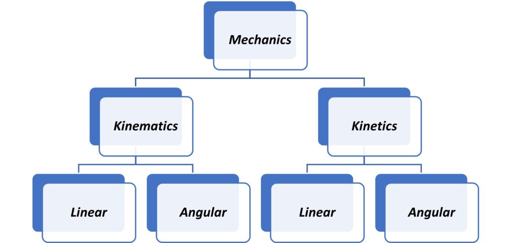
 

Figure1. The subdivisions of mechanics explore position, velocity and acceleration

Mechanics
Mechanics is a field of study in the area of physics. It focuses on the effect of forces acting on a body. A force is basically a push or a pull applied to a body that wants to make it move. Mechanics looks at how a body is affected by forces applied by muscle, gravity, and contact with other bodies.

In the case of a horse in motion, the term “body” could be used to describe the whole horse or could also be used to describe an individual body segment such as the limb, digit, or foot, or even an individual bone within a segment. The hoof capsule is a three dimensional multi-directional reinforced composite structure and its shape and morphology are influenced by the opposing forces its interaction with the ground (ground reaction force) and descending body weight acting on it both during statically and during locomotion. The external shape of the hoof capsule reflects the distribution and magnitude of the stresses and strains that occur in the tissues and structures of the hoof during weight bearing and locomotion. Individual conformation will influence the orientation of the distal limb segments and as such is fundamentally related to the bio-mechanics of the hoof and its ability to distribute and dissipate and absorb forces. To study these forces the mechanical structure of the foot and its associated anatomical structures three types of mechanical behaviour can be explored in both a static and dynamic state.

Angular kinematics
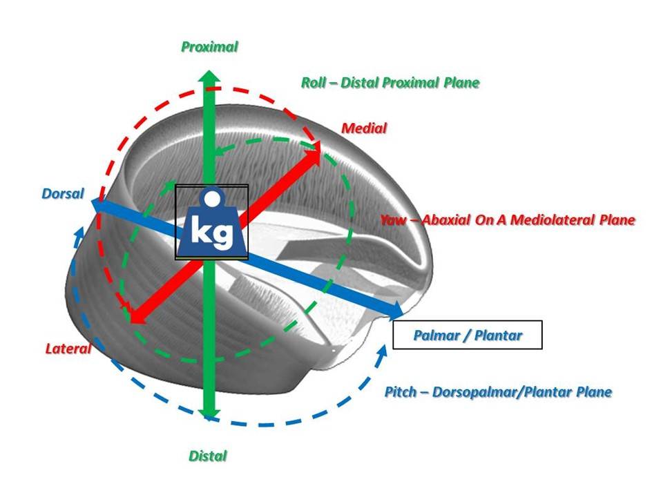
Angular kinematics describes angular motion, or motion involving rotations. Angular kinematics are used to describe the pitch, yaw and roll of the foot, Angular distance and angular displacement describe how far a body rotates. Similar to linear kinematics, angular distance means how far a body or segment rotates, while angular displacement means how far it rotates in a specified direction. Angular speed is just how fast the body rotates, but angular velocity refers to how fast it rotates in a specific direction.


FIGURE 2. Angular kinematics are used to describe the pitch, yaw and roll of the foot

Kinetics
Is the subdivision of mechanics focused on the forces that act on a body to cause motion. Basically, a force is a push or a pull exerted by one body on another body. A force, such as a push or a pull, can’t be seen only the effect of a force on a body is visible. An applied force wants to change the motion of the body — to speed it up or slow it down in the direction the force is applied.

Sir Isaac Newton formulated a set of three laws describing the cause–effect relationship between the force applied and the changing motion, or acceleration, of a body. These three laws are the foundation for using kinetics to analyse both linear and angular motion. The application of basic mechanics and Newton’s laws of motions the comparison of results of ground reaction force studies in the vertical axis (hoof capsule distortion) with results of ground reaction force in the horizontal axis (hoof or shoe wear) should make it possible to identify the relationship between both forces.

Newton’s Laws of Motion

1. A body continues in a state of rest, or of uniform motion in a straight line, unless or until acted upon by a force.
2. The acceleration of a body is proportional to the force acting upon it, and takes place along the line of action of the said force.
3. Every force induces an equal and opposite reaction.

This first law defines the term Force. Force is something which, by its self, produces acceleration. No earthly body ever observed has a “natural” motion unaffected by force.

The second law is concerned with proportionality i.e. if a horse moves at 2 meters per second, we can state with confidence that the distance travelled is proportional to the time taken the acceleration of the horses body is proportional to the force acting on it.

The third law is probably the most difficult to deal with, when a force exists between two separate bodies A and B, the force exerted upon B by A is exactly equal to the force exerted upon A by B but opposite in direction. Newton’s third law makes the assertion that the Earth is being pulled up by your body with the same force that the Earth pulls you down to it (Gravity).

Linear kinetics
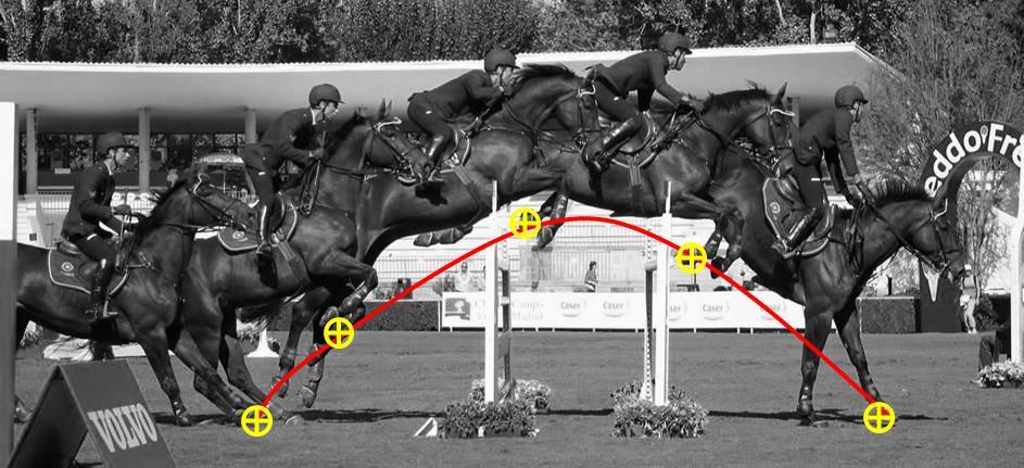
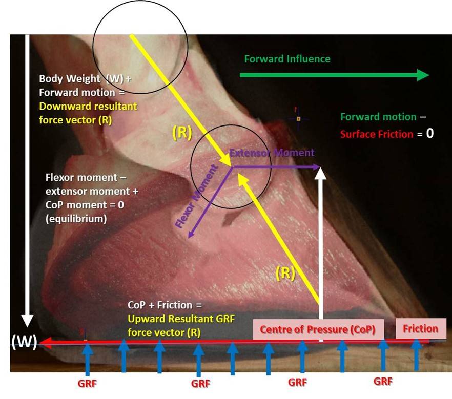
Linear kinetics investigates how forces affect the linear motion, or translation, of a body. The characteristics of a force include its size, direction, point of application, and line of action. Each characteristic influences the force’s effect on the body, and identifying the characteristics of each force applied to a body is an important step in kinetics. Typically farrier’s use the explanation of static equilibrium (figure 3) to describe the characteristics of a force and explain what makes gravity pull and friction push. A horse’s body during movement is usually acted on by several different external forces. The acceleration of the body is determined by the net force created by all the different forces acting at the same time creating what Newton describes as an unbalanced force.

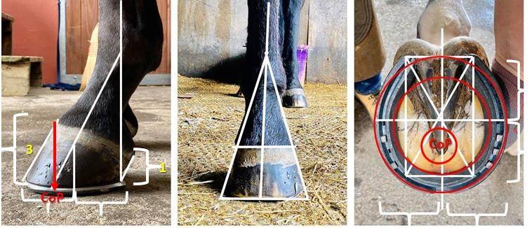
According to Rooney (1967) the vertical force of the ground reaction force (GRF) is exerted all over the bearing surfaces of the hoof in contact with the ground. In mechanics one considers that “spread-out” force to be concentrated at a single point called the centre of pressure (CoP). That is done in order to simplify the calculations. It does not mean that “all” the force is concentrated at that point; it means that one can account for the mechanics of the foot if one considers that the dispersed forces are all concentrated at that one point. Rooney gives good mathematical and diagrammatic explanations of the linear forces acting on the hoof that allow them to be plotted accurately (figure 3). Rooney also gives a simple definition of CoP as follows: that if a triangular support were to be placed under the horse’s foot at the centre of pressure, then at mid stance the well conformed and geometrically proportioned foot (figure 3) would not tip forwards or backwards but balance.

FIGURE 3. Each linear characteristic influences the force’s effect on the body, and identifying the characteristics of each force applied to a body is an important step in kinetics. Typically farrier’s use the explanation of static equilibrium
FIGURE 4. A well balanced foot – if a triangular support were to be placed under the horse’s foot at the centre of pressure, then at mid stance the well conformed and geometrically proportioned foot

Angular kinetics

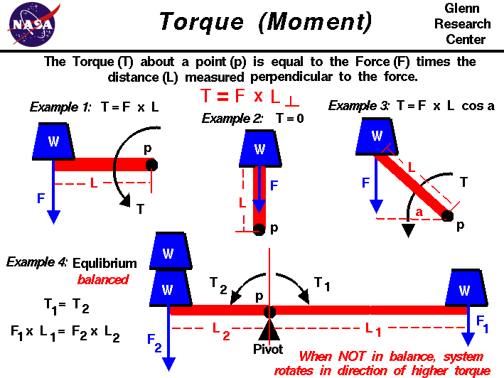
Angular kinetics investigates the causes of angular motion, or rotation. The turning effect of a force applied to a body is called torque. Torque is produced when a force is applied to a body at some distance from an axis of rotation (figure 5).

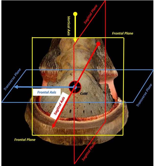
To describe movement, bio mechanists use a spatial reference system. All three dimensional structures, such as a horses foot, have three axis each of which has two coordinates. The three axes intersect at right angles to each other at the body or body segments centre of mass or balance point (CoP).The origin is at the intersection of the horizontal, mediolateral and vertical axes and is designated as zero (0) (figure?).

FIGURE 5. this diagramatic explanation of torque demonstrates that A force may be thought of as a push or pull in a specific direction. When a force is applied to an object, the resulting motion of the object depends on where the force is applied and how the object is confined. If the object is unconfined and the force is applied through the center of gravity, the object moves in pure translation, as described by Newton’s laws of motion.

Standardizing a Reference Frame

To understand the nature of torque and how it affects the foot we must first use a standardised reference framework that applie to either the whole horse’s body or the individual segments such as the digit can move in many directions, creating a need to standardize the description of movement. Figure 6 shows the three standard planes and axis of the foot commonly used to describe magnitude and direction of movement.

A) Sagittal plane: The sagittal plane divides the body into right and left parts. When you look at foot from the side, you see it in the sagittal plane.
B) Frontal plane: The frontal plane divides the body into front (anterior) and back (posterior) parts. When viewing mediolateral hoof balance either from in front or behind someone, you looking through the frontal plane.
C) Transverse plane: The transverse plane, sometimes called the horizontal plane, divides the body into an upper (superior) and a lower (inferior) part. When looking at the solear border or looking down over the coronary border of the foot is being viewed in the transverse plane.

The planes are aligned at right-angles to each other. When a standard plane passes through the centre of gravity of the body it’s called a cardinal plane. The centre of gravity of the body or segment being studied is called the centre of mass which would be the balance point of the body if suspended in a free body diagram (no force acting upon it). A diagonal plane is a plane cutting across the cardinal planes at an angle. There are an infinite number of ways an oblique plane can cut through a body and these are described in common directional terms.

FIGURE 5. Planes of reference for the equine digit
FIGURE 7. Forelimb Ppaines of reference

To be continued

This paper describes a method of treating cases of unilateral palmar/plantar laminitis using the Steward Clog ®(aka Wooden Shoe). This method varies significantly from a previously advocated technique using the Wooden Shoe. In this paper we report on the use of a different/modified technique to load the unaffected wall in horses with marked unilateral displacement (including sheared heel) of the distal phalanx. Additionally, the EVA/Wood Clog used- represents further development of the shoe as originally described (Steward Clog 2.0®).
Introduction:

The variety of benefits the Steward Clog® (Wooden Shoe) offers the laminitic horse is evident by the widespread usage throughout the horse industry. Ease of application and design modifications allow the shoe to be utilized effectively by practitioner and/or farrier without an expertise in therapeutic shoeing. The application process allows a non-traumatic procedure that relies heavily on the input of the horse in maximizing the horse’s comfort. Lateral and dorsopalmar (DP) radiographs are vital information to aid the proper trim in the therapeutic shoeing procedure.

Unilateral distal displacement can be accompanied by dorsal capsular displacement, in complicated laminitic cases. The damaged lamellae allow overload injury to portions of the wall / bone interface and this can manifest itself in variable displacement of the distal phalanx within the hoof capsule. The mechanical pull of the deep digital flexor tendon, combined with the weight forces produce the most common manifestation of laminitis- dorsal capsular displacement/phalangeal displacement. The pattern of displacement varies with the distribution of damage and load, the exact pathophysiology of which has yet to be determined. Displacement of a particular portion of the third phalanx is usually due to the area of displacement being overloaded and the damaged Suspensory Lamellar Apparatus’ (SLA) inability to support the load. The classic manifestations of laminitis are usually manifested as overload injury of a particular area of the distal phalanx. Depending on the extent of lamellar damage and the particular amount of load to P3, the variable displacement of the third phalanx is indicative of amount of damage and amount of load causing shear forces to the SLA.

Unilateral displacement is usually medial in the front limbs and lateral (author’s opinion) in the hind limbs (see Fig.1) -in typical cases. Conformational imbalanced (valgus) overloading of the medial wall’s damaged lamellae (in the fore limbs- with non-ambulatory perfusion deficits) is thought to be the usual cause of fore limb’s medial displacement (see Fig. 2). The hind limb is (typically) imbalanced overloaded on the lateral aspect of the hoof (because of the single leg resting stance) and this can often be manifested as lateral displacement (if the lamellae have been sufficiently damaged and overloaded). The diagnosis is based on the physical appearance of the foot, asymmetrical pain distribution and radiographs. Dorsopalmar (DP) radiographs are very helpful in revealing the condition. Abscesses and other pathologies causing particular areas of vascular discrepancies can alter the common areas of manifestations of this pathology. Current dynamic Positron Emission Tomography (Florodeoxyglucose) scans (Andrew VanEps, DVM) have shown extensive areas of (ie,-medial) wall that have deficits in vascular perfusion when non-ambulatory- in clinically normal feet. These deficits resolve upon ambulation. This may account for the medial UPD when dorsal phalangeal laminitis occurs, for example. The non-ambulatory lesions become pathological when the patient suffers lamellae damage and the painful condition further diminishes the compromised SLA wall perfusion.

In cases of dorsal phalangeal rotation of the third phalanx, the anterior ½ of the coffin bone distal displaces around the second phalanx’s distal condyles. The caudal ½ of the coffin bone displaces upward (unless a sinker). The lamellae in the heel area are stretched and are receptive to weight load, which would apply forces to aid in realigning the lamellae as the weight forces the bony column (P3) distally and the ground reaction forces (GRF) produce an upward vector force on the frog area of the hoof. When the lamellae are sufficiently damaged and overloaded, shearing of the lamellae occurs and the weight forces (WF) cause the distal migration of the affected portion of P3 (ie. – medial wing). Heel hoof growth can be considered to be enhanced because of the wide growth rings, but most of the enlarged ring is due to lamellar stretching and the pull on the newly formed hoof wall tubules in an unencumbered growing environment just below the dorsally displaced coronet. The horn tubules may be formed at the same rate around the coronet, but the stretching/compression and traffic jam/ fast-lane areas of tubular maturation and development just below the coronet account for wall formation/growth and growth ring conformation / thickness. The dorsoaxially displacing wall helps to shear the SLA as the third phalanx displaces distally. This is a tectonic plate-like biomechanical problem- comparable to an earthquake.

Once the displacement has occurred, a dip (recess) can be palpated in the integument immediately proximal to the wall on the affected side. The ungual cartilage/ wall relationship is displaced. Additionally, the toe and unaffected heel of the hoof capsule displaces dorsoaxially and rotates towards (yaw effect) the separated side (the plastic deformation usually occurs over time). This displacement (distal pitch of P3) and rotation (yaw of the hoof capsule) increase with time as the wall on the affected side shows little or no growth (due to disrupted blood supply in the damaged SLA) and the opposite side shows normal or increased growth. As the third phalanx rotates distally (pitch), the affected hoof wall is plastically displaced dorsoaxially (medial/roll (inversion roll), pitch-distally). This displacement is similar in pathophysiology to cases of “sheared heels” (traumatic/subclinical unilateral palmar laminitis (stretching) with dorsoaxial wall displacement), but the amount of overload shear trauma (and SLA damage) to produce heel (P3 &/or heel wall) displacement in the two cases- is substantially different. Both cases occur as a result of overload (shear or stretching) to the (damaged) lamellar apparatus of the particular area of the hoof. Laminitis cases have an enzymatic damage/weakening of the lamellae that lends itself to failure- if the lamellar damage is sufficient and overload exceeds this overload failure point. Unloading the affected wall is essential to the success of therapeutic intervention in both conditions. In severe cases of unilateral palmar/plantar displacement (UPD), extreme measures usually have to be implemented to have a chance of success. This involves unloading the affected wall by floating (cutting short/unloading) the affected heel, load redistribution, moment arm force reduction- to the affected lamellae, unloading pained areas, and stabilizing P3. The upward displacement of the hoof wall and distal displacing third phalanx create complicated biomechanics for resolution, as the wing of P3 is forced upward (biomechanically) as the coronet and affected wall needs to be displaced distally via plastically deforming biomechanics. The new growth attachments must be plastically deformed or artificially moved back into a more normal relationship- for functional healing to occur.

Previous descriptions¹ of the use of the Clog (wooden shoe) in the treatment of unilateral distal displacement have focused on extending the shoe towards the unaffected side to shift the center of pressure away from the most damaged lamellae, but the author finds this somewhat useful in some chronic, stable pathology, but not relevant in cases of acute laminitis. (See drawing-A.Parks)

The full roller motion design feature, particularly the mediolateral breakover (roll) portion of the Wooden Clog, and the subsequent use of an ethylene vinyl acetate (EVA) pad (similarly designed)- allowed the patients to (immediately) formulate a consistent, comfortable therapeutic formulation that enhanced soundness. This palliative effect, combined with other procedures (ie.- coronary grooving/wall resection) produced the most consistent success rate (curative effect).

Materials and Methods:

Plywood (1.125 inch) is cut and shaped to form the basic Wooden Shoe design. Mediolateral sloping can be increased to allow the patient to easily manipulate foot loading. The longer the slope (D/P) is extended toward the centerline of the shoe, the easier mediolateral (roll) breakover is achieved (see Fig. 3) and the hoof can shift weight to the less painful heel by slightly rolling the shoe- inversion roll for medial UPD and eversion roll for lateral UPD.. The sloping can be extended past the centerline to form a wedge effect to the shoe with the widest portion of the wedge under the affected heel (see Fig. 4).
The addition of EVA (ethylene vinyl acetate) to a layer of plywood (with the perimeter of the EVA cut in the same shape as the basic Wooden Shoe- Steward Clog 2.0®) allows the patient to self-adjust (plastically deform) the EVA and adds the same biomechanics to the shoe (see Fig. 5). Additional concussion absorption is a very beneficial feature- as well as the selective stabilization the elastic, plastic properties the EVA possesses.

Results:

Five horses (Four QH, 1 Arabian, ages 3 – 20 years) with bilateral medial displacement (front feet), 9 cases (8 QH, 1 Paint, ages (2-22 years) with unilateral displacement (seven front and 2 hind feet) have been treated using this technique. Increased comfort and a better radiographic DIP joint alignment were noted when modifications were made.
Successful outcome depended on the amount of lamellar damage and the amount of vascular damage as evidenced by bone loss and permanent bony displacement. Four horses (4) with minimal bilateral displacement- returned to pasture sound, six (6) patients with minimal unilateral displacement in a single foot are (occasionally) ridden at a walk. Four (4) of the horses are maintained in restricted environments with limited soundness.
Six feet required partial wall resection after displaying wall detachment (greater than 1 inch of detachment). The (dead / nutritionally compromised) wall below the attachment appears to shrink (dehydrate) toward the distal phalanx and the growth from the coronet prolapses over the distal wall if it is not removed. The wall detachment is often mis-diagnosed as a “gravel abscess” as it presents itself at the coronary band.

Discussion:

This shoe allows the patient to selectively load and self adjust breakover (pitching) and wedging (roll). The Steward Clog (aka Wooden Shoe) requires the practitioner have a knowledge of the breakover needs of the particular case: whereby, mediolateral point of breakover (roll) is appropriately applied to a nondeformable shoe material (wood, steel, aluminum, rubber, etc.). The addition of EVA to the shoe’s solar surface allows the patient to apply the point of breakover- and subsequent wedging effects to the shoe. Both shoe designs enable the horse to re-adjust the mediolateral breakover as the hoof growth’s mediolateral plastic asymmetries occur between shoeings.

The sloping of the mediolateral surface to the (EVA/Wood, Wood) Clog allows the patient to “roll” the shoe to realign the DIP joint and to load the hoof according to the comfort of the patient. The use of the EVA material on the bottom of a layer of plywood allows the patient to easily, immediately conform the shoe to the biomechanical / comfort needs of the patient (Steward Clog 2.0®). The plastic properties of the particular EVA allow the basilar surface (usually toe in typical laminitis, but medial or lateral heel in cases of UPD-both deformations may be needed in a particular case) to maintain the plastically compressed areas. All the while, the EVA maintains elastic properties to absorb concussion and further conform as the hoof grows and horse heals- or pathology worsens. These cases- immediately- loaded the shoe to the unaffected side, wedging (mediolaterally) the shoe such that the DIP joint was realigned (see Fig. 1,3,5 ) when the EVA foam was present. This is analogous to how a person would walk if the suffered a cut to the medial heel of their foot. One would roll up (dorsodistally pitch) off the heel (inversion), load the lateral portion of the foot (heel) and place the foot towards the midline (axially) to walk (redistribute load). The base slopes of the shoe can be altered to redistribute load. The affected side’s basal shoe heel/wall/ toe solar edge can be beveled at a 35 degree and the unaffected (lateral shoe branch) side can have a lesser slope to the ground connected surface (0 to 15 degrees). This will transfer more load (GRF/WF) to the lateral wall.
The basal (ground) surface of the Wood Clog (or urethane Clog) can be modified by solar grinding or addition of a wedged pad to produce an inversion roll (wedge sloped laterally). This will painfully overload the medial wall; therefore, wall floating measures should be implemented and load redistributed to offload the affected wall’s/solar load share. The dorsal surface of the “wooden shoe” should be ground down starting at the second nail hole and gradually slope the wall and solar area caudally to the affected heel. The triangular shaped area is from the wall to the medial sulcus of the frog. The surface of the shoe will be removed at a continual deepening slope to unload the floating wall. The wall can be trimmed in a sloping fashion (opposite the sole recess) to insure wall contact is avoided. The dorsoaxially displaced wall/coronet will usually migrate distally- to return to a more normal position – if adequately unloaded. Another option is to construct a W shaped caudal pad to redistribute the load off the medial wall (see Fig.7). Leather or wood can be used to construct the W pad. It is extended dorsally to the quarters. The pad(s) are under the shoe, and extend to the caudal region to provide wall and frog support. The affected side can be reduced by grinding the shoe surface of the pad in the above described fashion from the 2nd nail hole to the heel. The recessed area will extend under the bars and extend to the medial sulci of the frog. The recess should extend forward to unload the area of the sole that would be occupied by the distally projected shadow of the wing of the P3. The central leg of the W is to support the frog. The pad can be expanded in the frog area to accept more load by adding a layer of SIM to the central and lateral sulci of the frog. A configured pad (thickened frog) can be used to make sure the frog is actively loaded to overload the frog and make sure the medial wall and sole are protected (unloaded). Wetting a leather pad insures proper fit once the pad is applied and allows easier plastic deformation- via hammering. The pad can be conformed (hammered) on an anvil – (much the same as steel) to thin the medial wall/solar area- from the second hole, to the medial sulci of the frog and caudally to the heels ( wall to frog sulci / quarter to heel recess). This orthotic package (purchase) can be shod with a normal shoe that has the medial branch unloaded as described above. This is useful for a sheared heel that must continue working. A (Z bar) shoe can be used to transfer load to the lateral wall in some cases. The manipulative mixing, curing of the sole impression material can offload the affected heel and overload the unaffected heel. For example, the Shore value of SIM can be softened by 50% by using ½ the white portion of the 2 part SIM in the mix. This will reduce the Shore value by 50% and soften the SIM, effectively unloading pained areas. Curing/ manipulative adjustments can be used to overload normal areas of the frog/unaffected heel. Loading the hoof just prior to the SIM setting up, and not fully loading the leg will allow the unaffected area to be (actively) overloaded- increasing the area’s share of WF.

Excessive damage to the affected wall can result in an (comparatively) increase in hoof growth on the unaffected side as compared to the affected side. This causes increased wedging (pitch-d/p- and roll m/l) to the hoof capsule between trims (see Fig. 5). The affected wall (or portion) has lost a large portion (or all) of the nutritional and physical support of the SLA. This can cause a rotational hoof capsular deformity (yaw effect), over time, as the affected heel dehydrates and shrinks. Deleterious wall compression and detachment needs to be aggressively dealt with. Coronary grooving and other wall sculpting, floating methods (resections) must be done to allow the displaced wall and prolapsed coronet to return to a more normal form to re-establish a more normal function. These wall growth distortions cause a force on the elastic/plastic hoof capsule that results in the plastic yaw motion/deformity to be toward the affected side,. The faster growing un(less)affected side adds substantial mass to the lateral wall (in cases of medial sinkers) and further adds to the medial yaw effect. The dorsal capsular rotation of P3, especially if severe (incrementally -degreed), confounds the yaw effect. The rapid growing (net vector effect) heel causes a medial yaw and the slow growing toe region produces an initial, distally- orientated plane of growth (just distal to coronet) as it seeks the distal extensor process (SLA) of P3. The dorsoaxially displaced proximal wall and coronet compromises (via stretching or compression) the coronary vascular and needs to be encouraged to plastically return to a more normal architecture to return a more normally functioning wall. This encourages the mitotic area to restore the new portion (section) of connecting tubules as they encounter the previously produced, immature, (coronary band- terminal papillae produced/ very pliable) wall tubules, thus, distortions of growth rings occur. These distortions are a function of the amount of rotation that P3 has suffered. Their growth is retarded by coronary band vascular compression/stretching- because of P3’s displacement,’ thus denying them of the normal nutrition of the unaffected heel lamellar circulation. As the unkeratinized (uncemented, pliable, connected) terminal tubules (continuously) exit from their coronary papillar finger-like factories, they normally are attached (easily) to the SLA (suspensory lamellae apparatus).

The normal wall tubule is displaced distally as it is seeks to be attached to the SLA- in a complex manner- as to allow a ratcheting growth pattern of the continuous tubule/lamellae complex. The tubules are responsible for the observed growth rings of the hoof. In their pliable state, as they exit the coronet (papillae factory), they are easily deformed. The wall tubules are similar to hair shafts and the collective intercemented- collection of tubules- is the hoof capsule. They are interconnected to each other to form the continuous, complex wall -with a (normally) very tough hoof wall cementing system, -after their detoured route caused by P3 displacement. They can encounter previous (slow) growth which causes a “traffic jam” and results in very compacted, small growth rings, or the tubule can be folded resembling an accordion or “pleats”(displacement dependent). In severe P3 rotation (greater than 25 degrees), the large pleated sheets of (horizontally connected) tubules can appear as “folded layers” as they form unstable, distorted hoof wall. The interconnected (horizontally) tubules are connected to the (individual) continuous (coronet to ground) hoof capsular tubules as the individual tubules follow their previously produced segments to the ground. The keratinazation and “cementation” occur (only) within the perimeters of their close proximity to the lamellar corium (SLA). It is damaged by the displacement of P3, and often does not return to an acceptable / functional state, thus distorting normal wall formation and growth.

Unilateral palmar displacement is a very difficult, devastating overload injury / manifestation to cases of complicated dorsal capsular/ phylangeal displacement laminitis. The amount of damage to the entire lamellar interface¹ combined with the amount of load to a particular area of the lamellar interface accounts for the various manifestations of laminitis. Reducing load to the lamellae- prior to devastating overload injury- is possible in some cases, but unfortunately, some cases experience devastating lamellar damage and overload injury / consequences- such that therapeutic biomechanical manipulation aimed at lessening / redistributing the load is of no benefit in re-establishing acceptable long-term soundness.

The aggressive use of coronary grooving / affected wall resection and unloading the affected wall- by trimming to allow the coronet to unload and return to a normal position -is highly recommended by the author. Any laminitic episode in the palmar (plantar) area complicates healing due to the blood supply (hemodynamics) and load bearing features generally required by the heels.

Fig. 1. a,- Illustrated radiograph showing the radiographic changes on the DP in UPD, This hind limb suffered lateral (left) displacement of the distal phalanx as evident in this dorsopalmar radiograph (b.). The increased distance in the DIP lateral joint space (white line), increased thickness of the lateral hoof wall, and differential lines of the nutrient foramen of P3 compared to the distal aspects of P2- all suggest unilateral diplacement (a., b.).

Dr. Andy Parks’ sketch of the laterally placed shoe on this depiction of a medial sinker that was described the AAEP 2007 paper- How to treat 3 Manifestations of Lamimitis using the Wooden Shoe (O’Grady, Steward, & Parks).

Fig. 2. This acute laminitis case was suffering pre-existing mediolateral imbalance that was possibly creating increased loading to the medial lamellar interface. Both front feet of this bilateral valgus deviated horse were shod similarly to the above radiograph. Wooden shoes were applied and this hoof required medial hoof wall resection 45 days later (see Fig. 5) due to severe wall disruption. Note the abnormal radiolucent area (lamellar interface area) on the medial aspect (arrow).

Fig. 3. The hoof in Fig. 1 was shod using the wooden shoe. No trimming was done to the hoof between shoeing. The shoe has been outlined in white lines. The arrow shows the medial slope (red line) that was extended to past the shoe’s midline after extending the shoe medially. (The slope of the red line is exaggerated for illustration purposes.) Note how the joint space has realigned in a more normal manner. This is accomplished by allowing the horse to increase load to the medial heel in this lateral displacement. Hoof placement will be abaxially in lateral displacers, axially in medial displacers.

Fig. 4. Following separation of the medial wall at the coronet, the separated hoof capsule was resected. The growth rings proximally illustrate the disparity in hoof growth between the medial and lateral wall before resection; the lateral wall markedly outgrew the compressed medial wall. Prior to application of the shoe, the coronary band was tilted down towards the medial side. The foot is twisted (medial jaw inversion roll) to the medial side (red line) due to disparity in heel wall growth and mediolateral shoe positioning. Following application of the shoe, the EVA compressed such that the medial coronet is now proximal to the lateral coronet (inversion roll).

Fig. 5. This EVA / Wooden shoe has self-adjusted to meet the biomechanical and comfort needs of this medial sinker. The lateral heel has grown more than the medial heel and the shoe can readjust for this difference. Soundness improved in the 30 days this particular shoe was worn due to hoof stabilization and wall resection. The resected wall allows the upward displaced coronet to plastically return to a more normal architectural arrangement with P3.

Fig. 6. This drawing illustrates the loading and compression of the EVA material in this unilateral displacement drawing. The mediolateral wedging effect realigns the joint space and aids in comfort of the patient. The radiograph displays the joint and shoe angles. The sole was trimmed with no mediolateral imbalance 30 days prior. Note the upward displacement of the affected coronary band.

Fig.7. This leather “W pad” is used to redistribute weight to the palmar/plantar hoof. The affected side of the UPD case can be unloaded by thinning the leather pad on the affected side by “hammering the wet leather pad on the anvil” prior to application. Other methods- such as recessing the shoe or cutting (floating) the affected wall- can be used to offloaded the pained region of the hoof. The unilateral wing of the displaced third phalanx is usually very painful and warrants offloading with less sole impression material, or a softer durometer SIM in the pained areas. Usually a combinations of offloading are done to allow the new wall to regain a normal SLA attachment. Photo B. The affected side can be ground down to offload the sore solear area- the entire area from the quarter to the heel if needed.

Fig. 8 (a. b. c.) These pictures are of a healed medial UPD case that must be trimmed to offset the displaced medial wall (with chronic growth disparity). The upward shift of the medial wall and the distal displacement of the medial wing of P3 create trimming complexities and are distorted between trims due to wall growth disparities.

Fig. 9. (a. b. c.) These pictures show the proper trim and shoeing prescription provided by using an EVA Clog placed on the hoof. The conforming Clog plastically deforms to the special needs of these cases. Note the level coronet in picture a. (verses the same foot in Fig. 9a.) provided by the lateral compression of the Clog. The growth of the lateral wall promotes a medial jaw as the hoof grows. Shoeing allows the hoof to alter the hoof’s inversion roll as the lateral wall grows faster than the medial wall. The dorsal capsular displacement (pitch) is complicated by the medial unilateral palmar displacement (inversion roll) and will require special trims and shoeings for the life of the horse.

Acknowledgments:

i. Declaration of ethics- No ethics of the AVMA, AAEP, or AFA were violated in applications of these methods or writing this paper

ii. Conflicts of interests- The authors received compensation for the application and sale of the orthotics in treatment of these pathologies. The Steward Clog has been used extensively since 1986.

iii, Funding/Materials/Technical Support- No funding was provided by entities/businesses or material/technical support

References:

O’Grady, S.E., Steward, M., Parks, A.H. (2007a) How to Construct and Apply the Wooden Shoe for treating Three Manifestations of Chronic Laminitis. in Proceedings. Amer. Assoc of Equine Practnrs. 53, 423-429.

Steward, ML. How to construct and apply atraumatic therapeutic shoes to treat acute or chronic laminitis in the horse. American Association of Equine Practitioners 49th Annual Convention 2003;337-346.

Parks AH. Chronic Laminitis. In: Current Therapy in Equine Medicine. 2003 pp. 520-528. Saunders, Philadelphia.

Parks AH. O’Grady, SE, Chronic laminitis: Current treatment strategies, Vet. Clinics of N. America; 19: 393-416.

Steward, M.L. The Use of the Wooden Shoe (Steward Clog) in Treating Laminitis, Veterinary Veterinary Clinics of North America: Equine Practice- Laminitis, Vol. 26, Issue 1, April 2010, pages 207-214.

Hood DM. The mechanisms and consequences of structural failure of the foot
Vet. Clin. N Am 1999; 15: 437-461.

® Steward Clog (2.0)- Well Shod Farrier Supply, 305 N. Arthur St. Amarillo, Texas 79107 (806)322-3680 www. Well-shod.com