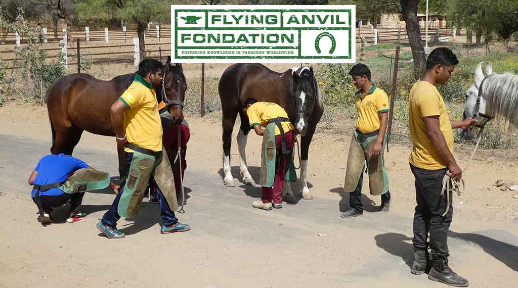
Introduction:
The variety of benefits the Steward Clog® (Wooden Shoe) offers the laminitic horse is evident by the widespread usage throughout the horse industry. Ease of application and design modifications allow the shoe to be utilized effectively by practitioner and/or farrier without an expertise in therapeutic shoeing. The application process allows a non-traumatic procedure that relies heavily on the input of the horse in maximizing the horse’s comfort. Lateral and dorsopalmar (DP) radiographs are vital information to aid the proper trim in the therapeutic shoeing procedure.
Unilateral distal displacement can be accompanied by dorsal capsular displacement, in complicated laminitic cases. The damaged lamellae allow overload injury to portions of the wall / bone interface and this can manifest itself in variable displacement of the distal phalanx within the hoof capsule. The mechanical pull of the deep digital flexor tendon, combined with the weight forces produce the most common manifestation of laminitis- dorsal capsular displacement/phalangeal displacement. The pattern of displacement varies with the distribution of damage and load, the exact pathophysiology of which has yet to be determined. Displacement of a particular portion of the third phalanx is usually due to the area of displacement being overloaded and the damaged Suspensory Lamellar Apparatus’ (SLA) inability to support the load. The classic manifestations of laminitis are usually manifested as overload injury of a particular area of the distal phalanx. Depending on the extent of lamellar damage and the particular amount of load to P3, the variable displacement of the third phalanx is indicative of amount of damage and amount of load causing shear forces to the SLA.
Unilateral displacement is usually medial in the front limbs and lateral (author’s opinion) in the hind limbs (see Fig.1) -in typical cases. Conformational imbalanced (valgus) overloading of the medial wall’s damaged lamellae (in the fore limbs- with non-ambulatory perfusion deficits) is thought to be the usual cause of fore limb’s medial displacement (see Fig. 2). The hind limb is (typically) imbalanced overloaded on the lateral aspect of the hoof (because of the single leg resting stance) and this can often be manifested as lateral displacement (if the lamellae have been sufficiently damaged and overloaded). The diagnosis is based on the physical appearance of the foot, asymmetrical pain distribution and radiographs. Dorsopalmar (DP) radiographs are very helpful in revealing the condition. Abscesses and other pathologies causing particular areas of vascular discrepancies can alter the common areas of manifestations of this pathology. Current dynamic Positron Emission Tomography (Florodeoxyglucose) scans (Andrew VanEps, DVM) have shown extensive areas of (ie,-medial) wall that have deficits in vascular perfusion when non-ambulatory- in clinically normal feet. These deficits resolve upon ambulation. This may account for the medial UPD when dorsal phalangeal laminitis occurs, for example. The non-ambulatory lesions become pathological when the patient suffers lamellae damage and the painful condition further diminishes the compromised SLA wall perfusion.
In cases of dorsal phalangeal rotation of the third phalanx, the anterior ½ of the coffin bone distal displaces around the second phalanx’s distal condyles. The caudal ½ of the coffin bone displaces upward (unless a sinker). The lamellae in the heel area are stretched and are receptive to weight load, which would apply forces to aid in realigning the lamellae as the weight forces the bony column (P3) distally and the ground reaction forces (GRF) produce an upward vector force on the frog area of the hoof. When the lamellae are sufficiently damaged and overloaded, shearing of the lamellae occurs and the weight forces (WF) cause the distal migration of the affected portion of P3 (ie. – medial wing). Heel hoof growth can be considered to be enhanced because of the wide growth rings, but most of the enlarged ring is due to lamellar stretching and the pull on the newly formed hoof wall tubules in an unencumbered growing environment just below the dorsally displaced coronet. The horn tubules may be formed at the same rate around the coronet, but the stretching/compression and traffic jam/ fast-lane areas of tubular maturation and development just below the coronet account for wall formation/growth and growth ring conformation / thickness. The dorsoaxially displacing wall helps to shear the SLA as the third phalanx displaces distally. This is a tectonic plate-like biomechanical problem- comparable to an earthquake.
Once the displacement has occurred, a dip (recess) can be palpated in the integument immediately proximal to the wall on the affected side. The ungual cartilage/ wall relationship is displaced. Additionally, the toe and unaffected heel of the hoof capsule displaces dorsoaxially and rotates towards (yaw effect) the separated side (the plastic deformation usually occurs over time). This displacement (distal pitch of P3) and rotation (yaw of the hoof capsule) increase with time as the wall on the affected side shows little or no growth (due to disrupted blood supply in the damaged SLA) and the opposite side shows normal or increased growth. As the third phalanx rotates distally (pitch), the affected hoof wall is plastically displaced dorsoaxially (medial/roll (inversion roll), pitch-distally). This displacement is similar in pathophysiology to cases of “sheared heels” (traumatic/subclinical unilateral palmar laminitis (stretching) with dorsoaxial wall displacement), but the amount of overload shear trauma (and SLA damage) to produce heel (P3 &/or heel wall) displacement in the two cases- is substantially different. Both cases occur as a result of overload (shear or stretching) to the (damaged) lamellar apparatus of the particular area of the hoof. Laminitis cases have an enzymatic damage/weakening of the lamellae that lends itself to failure- if the lamellar damage is sufficient and overload exceeds this overload failure point. Unloading the affected wall is essential to the success of therapeutic intervention in both conditions. In severe cases of unilateral palmar/plantar displacement (UPD), extreme measures usually have to be implemented to have a chance of success. This involves unloading the affected wall by floating (cutting short/unloading) the affected heel, load redistribution, moment arm force reduction- to the affected lamellae, unloading pained areas, and stabilizing P3. The upward displacement of the hoof wall and distal displacing third phalanx create complicated biomechanics for resolution, as the wing of P3 is forced upward (biomechanically) as the coronet and affected wall needs to be displaced distally via plastically deforming biomechanics. The new growth attachments must be plastically deformed or artificially moved back into a more normal relationship- for functional healing to occur.
Previous descriptions¹ of the use of the Clog (wooden shoe) in the treatment of unilateral distal displacement have focused on extending the shoe towards the unaffected side to shift the center of pressure away from the most damaged lamellae, but the author finds this somewhat useful in some chronic, stable pathology, but not relevant in cases of acute laminitis. (See drawing-A.Parks)
The full roller motion design feature, particularly the mediolateral breakover (roll) portion of the Wooden Clog, and the subsequent use of an ethylene vinyl acetate (EVA) pad (similarly designed)- allowed the patients to (immediately) formulate a consistent, comfortable therapeutic formulation that enhanced soundness. This palliative effect, combined with other procedures (ie.- coronary grooving/wall resection) produced the most consistent success rate (curative effect).
Materials and Methods:
Plywood (1.125 inch) is cut and shaped to form the basic Wooden Shoe design. Mediolateral sloping can be increased to allow the patient to easily manipulate foot loading. The longer the slope (D/P) is extended toward the centerline of the shoe, the easier mediolateral (roll) breakover is achieved (see Fig. 3) and the hoof can shift weight to the less painful heel by slightly rolling the shoe- inversion roll for medial UPD and eversion roll for lateral UPD.. The sloping can be extended past the centerline to form a wedge effect to the shoe with the widest portion of the wedge under the affected heel (see Fig. 4).
The addition of EVA (ethylene vinyl acetate) to a layer of plywood (with the perimeter of the EVA cut in the same shape as the basic Wooden Shoe- Steward Clog 2.0®) allows the patient to self-adjust (plastically deform) the EVA and adds the same biomechanics to the shoe (see Fig. 5). Additional concussion absorption is a very beneficial feature- as well as the selective stabilization the elastic, plastic properties the EVA possesses.
Results:
Five horses (Four QH, 1 Arabian, ages 3 – 20 years) with bilateral medial displacement (front feet), 9 cases (8 QH, 1 Paint, ages (2-22 years) with unilateral displacement (seven front and 2 hind feet) have been treated using this technique. Increased comfort and a better radiographic DIP joint alignment were noted when modifications were made.
Successful outcome depended on the amount of lamellar damage and the amount of vascular damage as evidenced by bone loss and permanent bony displacement. Four horses (4) with minimal bilateral displacement- returned to pasture sound, six (6) patients with minimal unilateral displacement in a single foot are (occasionally) ridden at a walk. Four (4) of the horses are maintained in restricted environments with limited soundness.
Six feet required partial wall resection after displaying wall detachment (greater than 1 inch of detachment). The (dead / nutritionally compromised) wall below the attachment appears to shrink (dehydrate) toward the distal phalanx and the growth from the coronet prolapses over the distal wall if it is not removed. The wall detachment is often mis-diagnosed as a “gravel abscess” as it presents itself at the coronary band.
Discussion:
This shoe allows the patient to selectively load and self adjust breakover (pitching) and wedging (roll). The Steward Clog (aka Wooden Shoe) requires the practitioner have a knowledge of the breakover needs of the particular case: whereby, mediolateral point of breakover (roll) is appropriately applied to a nondeformable shoe material (wood, steel, aluminum, rubber, etc.). The addition of EVA to the shoe’s solar surface allows the patient to apply the point of breakover- and subsequent wedging effects to the shoe. Both shoe designs enable the horse to re-adjust the mediolateral breakover as the hoof growth’s mediolateral plastic asymmetries occur between shoeings.
The sloping of the mediolateral surface to the (EVA/Wood, Wood) Clog allows the patient to “roll” the shoe to realign the DIP joint and to load the hoof according to the comfort of the patient. The use of the EVA material on the bottom of a layer of plywood allows the patient to easily, immediately conform the shoe to the biomechanical / comfort needs of the patient (Steward Clog 2.0®). The plastic properties of the particular EVA allow the basilar surface (usually toe in typical laminitis, but medial or lateral heel in cases of UPD-both deformations may be needed in a particular case) to maintain the plastically compressed areas. All the while, the EVA maintains elastic properties to absorb concussion and further conform as the hoof grows and horse heals- or pathology worsens. These cases- immediately- loaded the shoe to the unaffected side, wedging (mediolaterally) the shoe such that the DIP joint was realigned (see Fig. 1,3,5 ) when the EVA foam was present. This is analogous to how a person would walk if the suffered a cut to the medial heel of their foot. One would roll up (dorsodistally pitch) off the heel (inversion), load the lateral portion of the foot (heel) and place the foot towards the midline (axially) to walk (redistribute load). The base slopes of the shoe can be altered to redistribute load. The affected side’s basal shoe heel/wall/ toe solar edge can be beveled at a 35 degree and the unaffected (lateral shoe branch) side can have a lesser slope to the ground connected surface (0 to 15 degrees). This will transfer more load (GRF/WF) to the lateral wall.
The basal (ground) surface of the Wood Clog (or urethane Clog) can be modified by solar grinding or addition of a wedged pad to produce an inversion roll (wedge sloped laterally). This will painfully overload the medial wall; therefore, wall floating measures should be implemented and load redistributed to offload the affected wall’s/solar load share. The dorsal surface of the “wooden shoe” should be ground down starting at the second nail hole and gradually slope the wall and solar area caudally to the affected heel. The triangular shaped area is from the wall to the medial sulcus of the frog. The surface of the shoe will be removed at a continual deepening slope to unload the floating wall. The wall can be trimmed in a sloping fashion (opposite the sole recess) to insure wall contact is avoided. The dorsoaxially displaced wall/coronet will usually migrate distally- to return to a more normal position – if adequately unloaded. Another option is to construct a W shaped caudal pad to redistribute the load off the medial wall (see Fig.7). Leather or wood can be used to construct the W pad. It is extended dorsally to the quarters. The pad(s) are under the shoe, and extend to the caudal region to provide wall and frog support. The affected side can be reduced by grinding the shoe surface of the pad in the above described fashion from the 2nd nail hole to the heel. The recessed area will extend under the bars and extend to the medial sulci of the frog. The recess should extend forward to unload the area of the sole that would be occupied by the distally projected shadow of the wing of the P3. The central leg of the W is to support the frog. The pad can be expanded in the frog area to accept more load by adding a layer of SIM to the central and lateral sulci of the frog. A configured pad (thickened frog) can be used to make sure the frog is actively loaded to overload the frog and make sure the medial wall and sole are protected (unloaded). Wetting a leather pad insures proper fit once the pad is applied and allows easier plastic deformation- via hammering. The pad can be conformed (hammered) on an anvil – (much the same as steel) to thin the medial wall/solar area- from the second hole, to the medial sulci of the frog and caudally to the heels ( wall to frog sulci / quarter to heel recess). This orthotic package (purchase) can be shod with a normal shoe that has the medial branch unloaded as described above. This is useful for a sheared heel that must continue working. A (Z bar) shoe can be used to transfer load to the lateral wall in some cases. The manipulative mixing, curing of the sole impression material can offload the affected heel and overload the unaffected heel. For example, the Shore value of SIM can be softened by 50% by using ½ the white portion of the 2 part SIM in the mix. This will reduce the Shore value by 50% and soften the SIM, effectively unloading pained areas. Curing/ manipulative adjustments can be used to overload normal areas of the frog/unaffected heel. Loading the hoof just prior to the SIM setting up, and not fully loading the leg will allow the unaffected area to be (actively) overloaded- increasing the area’s share of WF.
Excessive damage to the affected wall can result in an (comparatively) increase in hoof growth on the unaffected side as compared to the affected side. This causes increased wedging (pitch-d/p- and roll m/l) to the hoof capsule between trims (see Fig. 5). The affected wall (or portion) has lost a large portion (or all) of the nutritional and physical support of the SLA. This can cause a rotational hoof capsular deformity (yaw effect), over time, as the affected heel dehydrates and shrinks. Deleterious wall compression and detachment needs to be aggressively dealt with. Coronary grooving and other wall sculpting, floating methods (resections) must be done to allow the displaced wall and prolapsed coronet to return to a more normal form to re-establish a more normal function. These wall growth distortions cause a force on the elastic/plastic hoof capsule that results in the plastic yaw motion/deformity to be toward the affected side,. The faster growing un(less)affected side adds substantial mass to the lateral wall (in cases of medial sinkers) and further adds to the medial yaw effect. The dorsal capsular rotation of P3, especially if severe (incrementally -degreed), confounds the yaw effect. The rapid growing (net vector effect) heel causes a medial yaw and the slow growing toe region produces an initial, distally- orientated plane of growth (just distal to coronet) as it seeks the distal extensor process (SLA) of P3. The dorsoaxially displaced proximal wall and coronet compromises (via stretching or compression) the coronary vascular and needs to be encouraged to plastically return to a more normal architecture to return a more normally functioning wall. This encourages the mitotic area to restore the new portion (section) of connecting tubules as they encounter the previously produced, immature, (coronary band- terminal papillae produced/ very pliable) wall tubules, thus, distortions of growth rings occur. These distortions are a function of the amount of rotation that P3 has suffered. Their growth is retarded by coronary band vascular compression/stretching- because of P3’s displacement,’ thus denying them of the normal nutrition of the unaffected heel lamellar circulation. As the unkeratinized (uncemented, pliable, connected) terminal tubules (continuously) exit from their coronary papillar finger-like factories, they normally are attached (easily) to the SLA (suspensory lamellae apparatus).
The normal wall tubule is displaced distally as it is seeks to be attached to the SLA- in a complex manner- as to allow a ratcheting growth pattern of the continuous tubule/lamellae complex. The tubules are responsible for the observed growth rings of the hoof. In their pliable state, as they exit the coronet (papillae factory), they are easily deformed. The wall tubules are similar to hair shafts and the collective intercemented- collection of tubules- is the hoof capsule. They are interconnected to each other to form the continuous, complex wall -with a (normally) very tough hoof wall cementing system, -after their detoured route caused by P3 displacement. They can encounter previous (slow) growth which causes a “traffic jam” and results in very compacted, small growth rings, or the tubule can be folded resembling an accordion or “pleats”(displacement dependent). In severe P3 rotation (greater than 25 degrees), the large pleated sheets of (horizontally connected) tubules can appear as “folded layers” as they form unstable, distorted hoof wall. The interconnected (horizontally) tubules are connected to the (individual) continuous (coronet to ground) hoof capsular tubules as the individual tubules follow their previously produced segments to the ground. The keratinazation and “cementation” occur (only) within the perimeters of their close proximity to the lamellar corium (SLA). It is damaged by the displacement of P3, and often does not return to an acceptable / functional state, thus distorting normal wall formation and growth.
Unilateral palmar displacement is a very difficult, devastating overload injury / manifestation to cases of complicated dorsal capsular/ phylangeal displacement laminitis. The amount of damage to the entire lamellar interface¹ combined with the amount of load to a particular area of the lamellar interface accounts for the various manifestations of laminitis. Reducing load to the lamellae- prior to devastating overload injury- is possible in some cases, but unfortunately, some cases experience devastating lamellar damage and overload injury / consequences- such that therapeutic biomechanical manipulation aimed at lessening / redistributing the load is of no benefit in re-establishing acceptable long-term soundness.
The aggressive use of coronary grooving / affected wall resection and unloading the affected wall- by trimming to allow the coronet to unload and return to a normal position -is highly recommended by the author. Any laminitic episode in the palmar (plantar) area complicates healing due to the blood supply (hemodynamics) and load bearing features generally required by the heels.