Scientific Horseshoeing logo
Introduction to Gross Anatomy and Physiology of the Equine Hoof

Gross Anatomy and Physiology of the Equine Hoof Part 1

by | May 16, 2022 | 0 comments

Introduction to Gross Anatomy and Physiology of the Equine Hoof

The reader should bear in mind that this text is not intended to be a fully detailed text on the anatomy or histology. The intention of this chapter is to highlight those aspects of the limb’s structure and their relationships most integral to the hoof and its role in locomotion. Further detail on the anatomy and histology of hoof horn and the other structures of the digit can be found in the scientific papers cited within this chapter.

The necessity for the study of anatomy
A farrier needs to have a good working knowledge of the anatomy (structure) and the physiology (function) of the horse and a detailed knowledge of the limbs of the equine particularly the lower limbs including the carpus (knee) and tarsus (hock). Because he/she is dealing with a living structure and his/her work can and does affect the performance and wellbeing of the whole animal he/she should also have a thorough working knowledge of the overall musculoskeletal unit.

The structure and function of the foot
The foot and hoof have three main functions, shock absorption, stabalisation and support for the musculoskeletal structure at rest or during motion and finally to facilitate movement. A high percentage of lower limb lameness problems either occur within or originate from the foot. Many of these lamenesses are often associated with continuous or repetitive strain caused bio biomechanical dysfunction. It is incumbent on every farrier should be aware of the anatomy, physiology and biomechanical functions of all structures within the foot enabling him or her to achieve the optimum biomechanical efficiency of the foot and limb and to have the confidence when dealing with both normal and abnormal foot conformations and associated lamenesses and pathologies.

A fundamental principle of farriery is to achieve equilibrium of static and dynamic forces acting on internal structures within the foot and lower limb. External influences should be neutralised by internal forces with the form of the foot maintained so as not to inhibit its natural function. Farriery attempts to maintain equilibrium within the foot by trimming to achieve an ill-defined and subjective empirical interpretation of “Static Foot Balance” (Hickman & Humphrey 1987; Stashak 2002). This interpretation of foot balance is mostly derived from historical texts (Dollar & Wheatley 1898; Russell 1897; & Lungwitz 1891). Today’s modern domesticated horse is far from the forces of nature that historically shaped and controlled their development. Horses are more reliant than ever on the knowledge and skill of the hoof care professional with an understanding of the anatomy, physiology and function of the lower limb, foot and all its component parts. The composition, position and functional relationship of the component parts of the foot are so densely compacted that within the hoof capsule there is little room for manoeuvre.

In nature shape and form of a structure relate directly to function. The hoof capsule is such an example of this concept. A horse’s natural protection against predators is “fright and flight” the hind quarters of the horse are a compact mass of large loco motor muscles which are designed primarily to propel the horse quickly forward. The front limbs are designed to primarily support the horse through impact, deceleration and loading during locomotion (Back & Clayton 2001). Movement is effected via neurological impulses causing the limbs to retract or protract. To minimise the energy required for this movement the limbs are as light weight as practical and are manoeuvred via a series of levers and pulleys. The pulleys are the joints, which are held together with a complex array of ligaments. The levers are the tendons which, together with the ligaments, act like a coil spring releasing stored energy when movement is necessitated. In simple terms every time the limb is loaded the elasticated collagen fibres that make up the tendons and ligaments store energy which is released when the limb needs to be lifted and propelled forward.

Anatomical Considerations
The gross anatomy of the foot and limb is well documented. In farriery the foot of the horse is referred to as the keratinized hoof capsule and its contents (figures 16 & 17). The hoof capsule is a continuation of modified epidermal tissue which forms a firm yet flexible protective layer surrounding the skeletal components at the distal extremity of the limb (Kempson. 1987, Dyson, 2011). The primary functions of the hoof capsule and its associated structures are to absorb impact shock during locomotion, assist in the transfer of weight from the skeletal column and protect the underlying structures whilst providing grip during locomotion (Butler 2005).

Each epidermal structure has a different density and hardness dependent upon its primary function and is produced by its own corresponding dermal layer and gains its strength and flexibility from its chemical composition and moisture content. The outer layer of hoof wall is rich in disulphides giving additional strength. The sole and frog are rich in sulfhydryl a group which gives these structures greater elasticity (Pollitt 1988) which gives flexibility. The average growth rate of 6-8mm per month (Stashak 2002) is said to be equal to the wear of the foot at the bearing border ground interface (Back and Clayton. 2001).
The hoof is the ultimate focus of forces generated by the limbs’ dual functions of weight bearing and locomotion. It is the only part of the horse’s anatomy whose shape, and consequently functions, can be altered readily. Hoof shape can be changed by the way those forces are directed into it (the way a horse moves), by weakening effects of environment, and by trimming and shoeing practices. Conversely, altering hoof shape can alter its functions and thus effect the movement and health of the limb above it. Because of this complex dynamic relationship between hoof shape, locomotion, environment and hoof care, detailed knowledge of hoof structure and function is essential for the practices of farriery and lameness related equine medicine.

The horse hoof is a horny covering on the end of each limb’s digit. Its purpose is in providing the limb with traction and protection. All such horny appendages are formed of keratinized protein. Among quadruped mammals, most have from two to five digits on the end of each limb that comprises their toes. Those mammals with two toes (deer, cattle, etc.) are termed “cloven hoofed.” The third phalanx of each of those two digits is encased in a hard, horny shell shaped something like a crescent to half circle. Together these two “claws” form a roundish base of support for the limb. The horse, however, has only a single digit on the end of each limb and thus its hoof is a single, solid horny case covering its single third phalanx. This single hoof distinctly resembles the two halves of the cloven hooves of larger two-towed quadrupeds “fused” together. Though these single and double digits are markedly different, they both must fulfil similar functions ultimately; supporting body weight and providing for locomotion while dissipating the shock and compression of these efforts. 

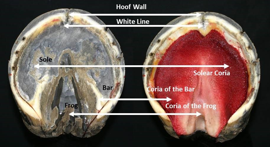
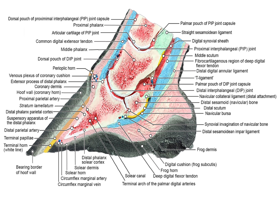
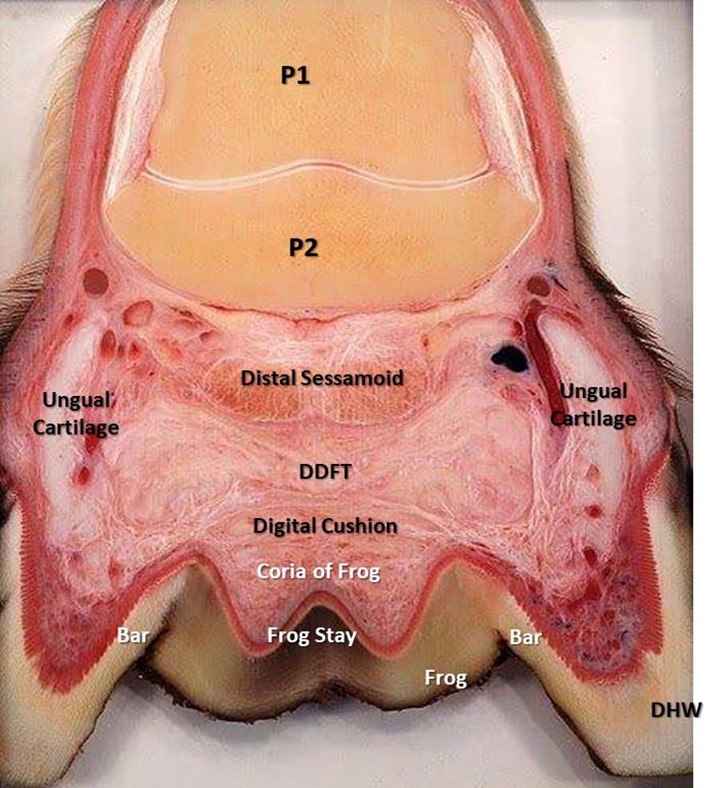
The hoof capsule encapsulates the structures of the foot including the distal interphalangeal joint (DIP joint), distal phalanx (P3), distal sesamoid (navicular) bone, dermal laminae, collateral ligaments, cartilages of P3, digital cushion, termination of the deep digital flexor tendon (DDFT) and a network of arteries, veins and nerves (Figure 2). The external bearing surface is comprised of the horny sole, white line and frog (Stashak, 2002). The hoof wall consists of three layers; the stratum external, stratum medium and the stratum internum (Stump, 1967, Reilly et al., 1996) The inner layers of the hoof wall, the stratum internum, consists of around 600 non-pigmented keratinised, primary epidermal laminae, each of which bears 100-150 non-keratinised, secondary epidermal laminae (SEL) (Stump, 1967). Pollitt (2001) confirmed that the SEL dovetail with their adjacent counterparts of the secondary dermal laminae (SDL) of the laminal corium (Figure 3), which covers the parietal surface of P3, suspending P3 within the hoof capsule. Between the dermal epidermal laminae there is a thin epithelial cellular layer, described as the basement membrane (BM), which undergoes constant remodelling (Pollitt, 2001). As a result the SEL slide past the SDL by breaking and reforming in a staggered ratchet-like manner so that the keratinised cells can move distally yet still support load (Pollitt, 2004).

Schematic diagrams showing the main anatomical structures of the equine foot in the midline sagittal plane (illustration courtesy of C. Pollitt.

The Component Parts of the Hoof

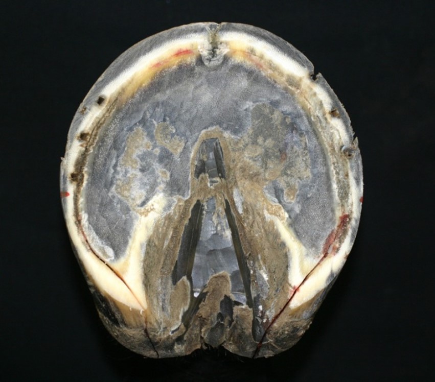
The insensitive horny hoof or hoof capsule consists of four basic component parts. These are the “wall,” the “bars,” the “sole” which covers most of the hoof’s lower or volar surface, and the wedge-shaped “frog” found on the volar surface between the bars and sole (figure 18). These parts, while produced by differing areas of sensitive tissues and composed if differing cell structures, are all fused together to form a single, flexible casing that is open at the top. Growth rates and amounts of horn in each hoof part can vary considerably relative to each other and between individuals.

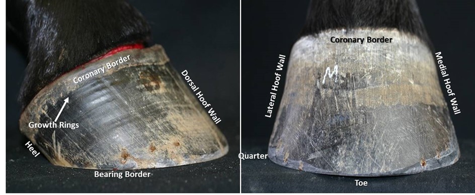
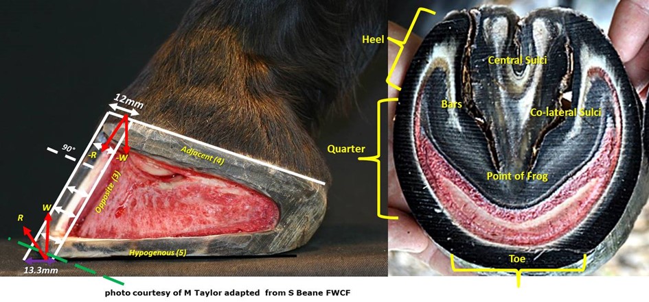
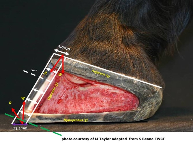
The hoof wall is the vertical structure arranged around the outer surface of the third phalanx, sloping forward and, to some degree, outward from the hairline region knows as the coronary band or border of the hoof. The wall is constructed of vertical tubules which are said to be parallel to the anterior surface of the third phalanx, the angle at which weight is loaded onto the wall from the bone. The name coronary band is derived from the high percentage of blood vessels contained in it. The wall’s dorsal aspect is termed the hoof’s toe. The middle halves of its medial and lateral sides are called the hoof’s quarters or side walls. The posterior sections of the wall, beginning behind its widest point on each side, are termed its heels. That widest aspect of the side walls is known as the “bend of the quarters”. The heel region includes the end of the wall where it meets the bars. This angles juncture of wall and bar is called the buttress of the heel (figure 4). The wall itself is thickest, horizontally, at the centre of the toe where it is approximatly10mm to 12mm across. From that point it tapers to its thinnest points near the bend of the quarters. It then normally thickens as it becomes the heel and heel buttress. The wall serves primarily to support body weight transmitted through its connection to the third phalanx as well as provide traction where its bearing border interacts with the ground surface.

The bars are actually continuations of the wall itself, turned inward and forward from the buttresses of the heel. The space between the wall of heel and the bar just forward of the buttress is termed the angle of the wall. The bars have some vertical height, like the wall, which diminishes to nothing toward the point of the frog (figure 4). The bars are slanted at an angle to the ground similar to that of the wall, so that their upper borders inside the hoof are closer together than their lower one on its volar surface. But the vertical surfaces of walls and bars are not parallel. The bars serve to reinforce the outer wall of the hoof by bracing and anchoring the heels, and also act to support the frog between them.
The frog is a mass of rubbery horn situated between the buttresses and bars, tapering forward toward the toe. Deep grooves on either side of the frog, separating it from the bars, are referred to as the collateral sulci or the commissures. Its narrow apex end is known as the point of the frog (figure 4). The groove or depression on the frog’s palmardistal surface which divides it into two halves is its central sulcus, sometimes termed the cleft. On the upper or interior surface, inside the hoof, the central sulcus forms a protrusion known as the frog stay (figure 5). The frog is supported on either side by the bars to which it is fused and serves primarily as a protective pad for the sensitive parts contained in the hoof above it.

the wall itself is thickest, horizontally, at the centre of the toe where it is approximatly10mm to 12mm across. From that point it tapers to its thinnest points near the bend of the quarters. It then normally thickens as it becomes the heel and heel buttress
the central sulcus forms a protrusion known as the frog stay. Photo by Dr. Christoph von Horst

Above and palmar aspect of the frog is a region called the bulbs of the heels. These two bulges below the hairline, from which the buttresses of the heels originate, are a thin covering of horny material over the soft palmaproximal border of the digital cushion. The horn of the bulbs helps protect the palmar structures of hoof including digital cushion.

The horny sole is the region of the hoof’s volar surface bounded by the bearing surface of the hoof wall, the bars and the frog. It tends to be concave rather than flat, arching up into the curvature of the third phalanx’s distal surface (figure 20). The sole is attached to the inner border of the wall by a flexible strip of horn called the white line (figure 4 ) binding the sides of the wall together laterally across the hoof, as well as to the frog and bars at their common borders. The vertical thickness of the sole is normally between 10mm and 12mm. It serves to It also protects the sensitive tissue above it from injury.

The Hoof Parts as a Whole

The hoof as a whole serves two basic functions. 1) Its overall shape is a medium for support and traction in locomotion. 2) It provides protection to the sensitive parts within and above it by being a resilient covering and dissipating internal pressures through its elastic composition and expansive actions. These functions require conflicting properties of rigidity for support/traction and flexibility for shock dissipation. Both the design of the hoof’s overall shape and the material properties of the horn from which its parts are formed enable it to perform these dual functions effectively.

The hoof wall is extremely efficient at defusing shock vibrations generated when the hoof impacts the ground during locomotion. It is able to reduce both the frequency and maximal amplitude of the vibrations (Dyhre-Poulsen et al., 1984). By the time the shock of impact with the ground reaches the first phalanx up to 90% of the energy has been dissipated, mainly at the lamellar interface.

The overall shape of the hoof is dictated by the direction from which weight and force are exerted upon it. Since weight is transmitted downward and forward through the bones of the digit, the hoof grows so that its greatest strength is aligned with those bones (figure 2). The impact of weight is thus thrown upon the more rigid forward aspect of the hoof wall, while its descending forces and those of propulsive effort are dissipated by the more elastic structures situated in the “gap” between the ends of the hoof wall at the heels.

The bars direct any weight falling on their upper border out toward the heels and wall, while at the same time limiting the expansion of the heels to avoid their collapse. The frog accommodates the lateral spreading of bars, heels and quarters under pressure by stretching laterally itself. A vertical section through the palmar third of the hoof shows this lateral spring structure of wall, bars and frog (figure 5). For the expansion and thus protective actions to occur, there must be some vertical length bars and the wall in the heels. It is important to understand that individual hoof walls come in various degrees of flexibility and will deform in any direction when a sufficient force from any angle overcomes the limits of the horns elasticity, consequently we must understand the usual forces applied to a hoof capsule and its morphology during locomotion to get any idea of what is normal distortion. Also we must consider the effects of not only the ground’s reaction but the changes in weight bearing caused by the animal’s centre of weight gravity as it moves over the hoof. The effects of weight bearing during the stance phase will be discussed in detail in a subsequent post.


figure 6 the sole is concave rather than flat, arching up into the curvature of the third phalanx’s distal surface

Epidermal structures within the hoof

The Hoof Wall
It is generally accepted that the hoof wall does not vary in thickness from its origin at the coronary band to its conclusion at the distal border. The wall is thicker at the toe and becomes progressively thinner and more elastic toward the heels where it thickens again where it reflects dorsally as the bars. This variation in hoof wall thickness is believed to be of importance in facilitating hoof function (Douglas et al. 1996, Pollitt 1996, O’Grady 2009 and Hampson and Pollitt 2011). When viewed from the solar surface the thickness of the hoof wall, around the foot’s circumference, can be evaluated to determine how much hoof might be removed when trimming or shoeing. This view can be misleading as to the true thickness of the hoof wall because the wall has either been pared or worn parallel to the ground surface. This creates a misleading view and could lead to an inaccurate assessment and is largely due to the angulation of wear or trim occurring across the dorsal hoof wall instead of at a right angle (figure 7). Balchin (2012), in a study of the sagittal section of 200 cadaver front feet, demonstrated that the coronary band and DHW where at right angles and formed a right angled triangle with the DHW and bearing border. The DHW is said to be of uniform thickness when viewed in a transverse section from its origin at the coronary border to the ground bearing border and parallel to the dorsal surface of P3. However the BB of the DHW presents perpendicular to the axial skeleton and oblique to the coronary border and is therefore increased in width at the perpendicular ground-bearing surface (figure 21A). Numerous authors have identified the importance of a hoof wall gradient from toe to heel, from both an anatomical and engineering prospective. Moore (2017) concluded that the natural circumferential toe to heel gradient of 1.77:1 was less than previously thought. Beane (2018) supported this contention and gave consideration to differential trimming protocols of the DHW and demonstrated a significant difference in solar arch morphology where the DHW was reduced through dorsal wall rasping.

The hoof wall is made up of three layers: the stratum extremum, the stratum medium and the stratum internum. The stratum externum, or external layer of the hoof, consists of an extremely thin layer of tubular horn. It includes the periople, which covers the bulbs of the heels and a variable tubular horn. The remainder of the wall is covered by the stratum tectorium (also part of the stratum externum) which has a high fat content and so helps to prevent the movement of water through the hoof wall. It is this layer which given the un-rasped wall its shiny appearance.

The stratum medium, which comprises the bulk of the hoof wall, is composed of tubular and intertubular horn. It is this part of the wall which is produced by the coronary corium. In dark hooves, this layer is pigmented with melanin. Although there is a widespread belief amongst that white hooves are inferior to dark hooves, differences in the mechanical properties of pigmented and non-pigmented hooves have not been demonstrated. The stratum internum is the name given to the internal layer of the hoof wall and comprises the primary and secondary epidermal and dermal laminae. This region is always non-pigmented, even in dark hooves.

The horn itself is composed of densely packed, longitudinally aligned, horn tubules which are generated from tiny projections of the dermis known as papillae (figure 8). These tubules are cemented together by intertubular horn cells which proliferate from germinative cells of the dermis between the papillae.

The intertubular horn is formed at right angles to the tubular horn and gives the hoof wall a mechanically stable, multidirectional, fibre-reinforced composite (Bertram and Gosline, 1987). Interestingly hoof wall is stiffer and stronger at right angles to the direction of the tubules. This contradicts the usual assumption that the ground reaction force is transmitted proximally up the hoof wall parallel to the tubules. The hoof wall appears to be reinforced by the tubules but it is the intertubular material that accounts for most of its mechanical strength stiffness and fracture toughness. The tubules are three times more likely to fracture than intertubular horn (Leach, 1980; Bertram and Gosline 1986).

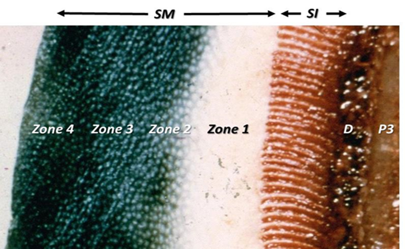
The bulk of the hoof wall consists of the stratum medium, which is the main load bearing part of the hoof wall, and extends from the coronary band (CB) to the bearing border (BB). Its generation is from the epidermal basal cells of the coronary corium (Stump, 1967; Pollitt, 2001) and it is a non-homogenous and anisotropic material within which horn tubes run diagonally from the CB to the BB. The horn tubules are arranged into four zones of density (Reilly et al. 1996), the strongest and most densely populated zone being the outer layer. Intertubular horn is formed at right angles to the tubular horn, filling the void between the horn tubules (Bertram & Gosline, 1987). Interestingly, hoof wall is stiffer and stronger at right angles to the direction of the tubules, a finding at odds with the usual assumption that the ground reaction force is transmitted proximally up the hoof wall parallel to the tubules. The hoof wall appears to be reinforced by the tubules but it is the intertubular material that accounts for most of its mechanical strength stiffness and fracture toughness. This construction achieves mechanical stability within the horn with the mechanical properties of the horn tubules being best suited to compressive force with the intertubular horn providing stability through tension (Bertram & Gosline, 1987). The equalisation of both compressive and tensile forces allows ground reaction forces to be dispersed within the structure without regional overload (Thomason, 2007). However a recent study (Lancaster et al., 2011) found a circumferential zonal pattern of tubule density with the medial and lateral locations showing the same radial pattern, but there was a significant difference in tubule density between the medial and lateral sides, and between the toe and the lateral quarter.

Figure 7 a schematic illustration of the DHW perpendicular relationship to the coronary border. The ground bearing border presented perpendicular to the force (-W) is increased by 10.83% increasing the moment. The resultant (R) GRFv induces a shear force which contributes to the dorsal migration of the dorsodistal hoof wall (flare). 
Figure 8 Hoof wall Intertubular material composite diagram. (A) A pixilated image of a 10 mm thick hoof wall cross-sectioned sample Surrounding the tubules are areas of Intertubular material, indicating a strong concentric organization of Intertubular intermediate filaments around the tubule cortex. (B) A three-dimensional representation of the Intertubular material. (C) Three-dimensional models of Intertubular material IF organization from each representative region. The orientation of Intertubular material varies depending on its position through the thickness of the hoof wall (Illustration with permission from A Parks modified after Kasapi and Gasoline (1997)).

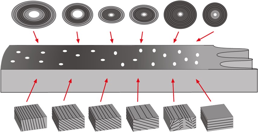
The highest tubular density is at the outermost layer (figure 9). Since the ground reaction force is transmitted proximally up the wall (Thomason et al 1992) the construction of the epidermal structures appears to be part of the mechanism that allows the transfer of impact load across the hoof wall from the rigid, high tubule density, outer wall to the more plastic, low tubule density, inner wall. The material properties of hoof are said to have plastic elastic characteristics (Reilly et al; 1996). These characteristics allow for deformation under stress and then return to original form following a limited period of strain. Maximum stress for prolonged periods of strain reduces the elastomeric property and the structures ability to return to its original form.

In a normal foot the entire hoof wall grows at a rate of approximately 6 to 10 mm per month. Growth is slower in excessively cold or dry environments. At the toe, it takes approximately 9 to 12 months for horn to grow from the coronary band to the ground surface, at the quarters 6 to 8 months, and at the heels 4 to 5 months. Because the distance from the coronary band to the ground is less at the heels than at the toe, heel horn at the ground surface is younger than toe horn, has higher moisture content and is more elastic. Significant changes in nutritional status, or persistent fever states, may cause changes in the growth of the hoof wall at the time of the “insult”. These become apparent as rings or ridges in the hoof wall, which then grow out with the hoof. The rings are usually roughly parallel to each other and to the coronary band. These should be contrasted with the rings on the hoof of a horse which has previously been affected by laminitis. “Laminitic” rings usually do not grown out parallel to each other because after laminitis as growth is often restricted at the toe resulting from disruption of the vasculature. Thus the ring is formed at the coronary band at roughly the same time all around the hoof, but the continued or accelerated heel growth causes sequential rings to diverge from each other at the heel as the hoof grows out.
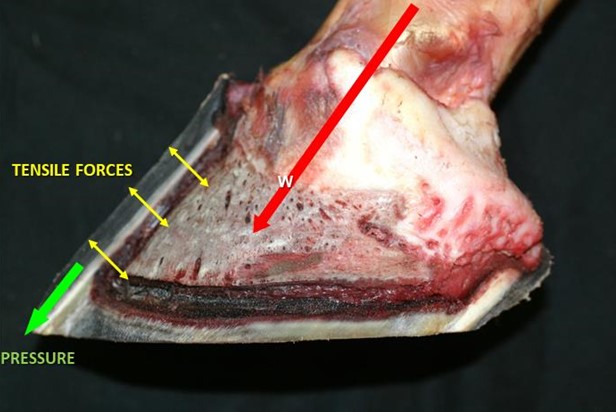
Similar to any other epidermal structure, it prevents absorption of water into the body from the environment and prevents loss of fluids from the body. It is effectively a “waterproofing layer”. But the function for which it is uniquely designed is that of weight bearing. The weight of the horse is transmitted down through the column of bones in the limb, ultimately being borne by the coffin bone.

Tightly adherent to the external surface of the coffin bone are the dermal laminae of the laminar corium. The tight interlocking of the dermal laminae with the epidermal laminae on the inside of the hoof wall allows the weight of the body to be transmitted via tensile force from the P3 to the hoof wall. Effectively the bodyweight is said to be “suspended” from the inside of the hoof, the ground reaction force being taken by the hoof wall.

There are some 600-900 primary laminae (figure 10) each with 100-200 secondary epidermal laminae (Pollitt. 1988). The primary epidermal laminae are produced by the dermis of the coronary corium. The secondary epidermal laminae are produced by a germinative layer of the epidermis of the laminar corium. The primary and secondary epidermal laminae interdigitate, dovetail, with the corresponding dermal lamellae. The dermal lamellae originate from the laminar corium surrounding the parietal surface of P3 and the lower border of the ungual cartilages. This relationship of interdigitation of laminae is thought to allow for the partial suspension of P3 within the hoof capsule and the transference of tensile forces radially from P3 (Thomason et al; 2009) (figure 10).

Figure 9 the inner layers of the hoof wall, including the stratum medium (SM), illustrating the differing layers of tubular density. The stratum internum (SI) consists of around 600 non-pigmented keratinised, primary epidermal laminae, each of which bears 100-150 non-keratinised, secondary epidermal laminae which dovetail with their adjacent counterparts of the secondary dermal laminae originating from the dermis (D). Modified after Rooney (1969).
Figure 10 the interdigitation of laminae is thought to allow for the partial suspension of P3 within the hoof capsule and the transference of tensile forces radially from P3 (Thomason et al; 2009)

In seeking to understand hoof function it is essential to grasp that the vast majority of weight is supported by the hoof walls through this lateral suspension from their inner border of the wall bears directly against the ground by the wall’s distal edge or bearing surface. The greater thickness of the wall toward the toe and the fact that the third phalanx is attached to the front half of the hoof wall indicate that more weight is to be supported there than by the thinner, less solidly anchored wall of the heels. Behind its midline, the hoof attaches via epidermal and dermal layers to flexible structures, such as the collateral cartilages, ill-suited to structural support. Further evidence of this dysfunctional dynamic of weight distribution is found in the declining density of primary lamellae in the wall as it approaches the heels (Bowker, et al; 2002). There are approximately 2.5 times more lamellae ridges per centimetre at the toe than at the heel. The greater density of lamellae at the toe indicates that this area provides greater support for weight. If the lamellae act primarily as slings holding the body weight in lateral suspension, they are subjected to constant tension, which increases with the speed of locomotion, except when the horse lifts a hoof or lies down. The consequences of excessive unnatural force distribution on the laminar connection are perhaps obvious.

In the normal foot, loading of the wall causes expansion of the hoof at the heels, and to a more limited extent at the quarter. Heel expansion is facilitated by the fact that heel horn is younger (and therefore moister) and thinner than the horn in other areas of the wall and so it is more elastic. This expansion presumably aids in diminution of the concussive forces associated with impact. Significant expansion therefore occurs only in those parts of the hoof wall which are not tightly bound to P3. There is evidence that the tensile strength of the laminae may actually lead to flattening of the hoof wall in the front part of the foot during weight bearing (Thomason et al; 2009). Shoeing has been shown to limit hoof expansion to some extent, apart from at the heels where the degree of expansion is unaffected, although the rate of expansion may increase.

How do we reconcile the fact that the epidermal hoof wall grows slowly towards the ground, retaining its tight weight-bearing bond with the underlying dermis, and yet the dermis remains in its original position adjacent to the coffin bone? Almost all cells are connected to their neighbours by a variable number of cell-to-cell junctions. These perform a variety of functions, such as allowing cells to communicate with each other, or forming an impenetrable barrier between the cells. There is a specialized type of junction called a desmosome which anchors cells to their neighbours, acting like a sort of intercellular rivet. The cells of the primary and secondary epidermal laminae are connected by desmosomes. It is thought that these desmosomes break and reform in a staggered sequence, allowing movement of the primary epidermal laminae over the secondary epidermal laminae, yet still retaining the strength of the laminar junction. This mechanism will be discussed in detail along with the functional anatomy of the remainig hoof structures in part 2 of this blog to published shortly.

Thank you for reading our article on Gross Anatomy and Physiology of the Equine Hoof.


This paper describes a method of treating cases of unilateral palmar/plantar laminitis using the Steward Clog ®(aka Wooden Shoe). This method varies significantly from a previously advocated technique using the Wooden Shoe. In this paper we report on the use of a different/modified technique to load the unaffected wall in horses with marked unilateral displacement (including sheared heel) of the distal phalanx. Additionally, the EVA/Wood Clog used- represents further development of the shoe as originally described (Steward Clog 2.0®).
Introduction:

The variety of benefits the Steward Clog® (Wooden Shoe) offers the laminitic horse is evident by the widespread usage throughout the horse industry. Ease of application and design modifications allow the shoe to be utilized effectively by practitioner and/or farrier without an expertise in therapeutic shoeing. The application process allows a non-traumatic procedure that relies heavily on the input of the horse in maximizing the horse’s comfort. Lateral and dorsopalmar (DP) radiographs are vital information to aid the proper trim in the therapeutic shoeing procedure.

Unilateral distal displacement can be accompanied by dorsal capsular displacement, in complicated laminitic cases. The damaged lamellae allow overload injury to portions of the wall / bone interface and this can manifest itself in variable displacement of the distal phalanx within the hoof capsule. The mechanical pull of the deep digital flexor tendon, combined with the weight forces produce the most common manifestation of laminitis- dorsal capsular displacement/phalangeal displacement. The pattern of displacement varies with the distribution of damage and load, the exact pathophysiology of which has yet to be determined. Displacement of a particular portion of the third phalanx is usually due to the area of displacement being overloaded and the damaged Suspensory Lamellar Apparatus’ (SLA) inability to support the load. The classic manifestations of laminitis are usually manifested as overload injury of a particular area of the distal phalanx. Depending on the extent of lamellar damage and the particular amount of load to P3, the variable displacement of the third phalanx is indicative of amount of damage and amount of load causing shear forces to the SLA.

Unilateral displacement is usually medial in the front limbs and lateral (author’s opinion) in the hind limbs (see Fig.1) -in typical cases. Conformational imbalanced (valgus) overloading of the medial wall’s damaged lamellae (in the fore limbs- with non-ambulatory perfusion deficits) is thought to be the usual cause of fore limb’s medial displacement (see Fig. 2). The hind limb is (typically) imbalanced overloaded on the lateral aspect of the hoof (because of the single leg resting stance) and this can often be manifested as lateral displacement (if the lamellae have been sufficiently damaged and overloaded). The diagnosis is based on the physical appearance of the foot, asymmetrical pain distribution and radiographs. Dorsopalmar (DP) radiographs are very helpful in revealing the condition. Abscesses and other pathologies causing particular areas of vascular discrepancies can alter the common areas of manifestations of this pathology. Current dynamic Positron Emission Tomography (Florodeoxyglucose) scans (Andrew VanEps, DVM) have shown extensive areas of (ie,-medial) wall that have deficits in vascular perfusion when non-ambulatory- in clinically normal feet. These deficits resolve upon ambulation. This may account for the medial UPD when dorsal phalangeal laminitis occurs, for example. The non-ambulatory lesions become pathological when the patient suffers lamellae damage and the painful condition further diminishes the compromised SLA wall perfusion.

In cases of dorsal phalangeal rotation of the third phalanx, the anterior ½ of the coffin bone distal displaces around the second phalanx’s distal condyles. The caudal ½ of the coffin bone displaces upward (unless a sinker). The lamellae in the heel area are stretched and are receptive to weight load, which would apply forces to aid in realigning the lamellae as the weight forces the bony column (P3) distally and the ground reaction forces (GRF) produce an upward vector force on the frog area of the hoof. When the lamellae are sufficiently damaged and overloaded, shearing of the lamellae occurs and the weight forces (WF) cause the distal migration of the affected portion of P3 (ie. – medial wing). Heel hoof growth can be considered to be enhanced because of the wide growth rings, but most of the enlarged ring is due to lamellar stretching and the pull on the newly formed hoof wall tubules in an unencumbered growing environment just below the dorsally displaced coronet. The horn tubules may be formed at the same rate around the coronet, but the stretching/compression and traffic jam/ fast-lane areas of tubular maturation and development just below the coronet account for wall formation/growth and growth ring conformation / thickness. The dorsoaxially displacing wall helps to shear the SLA as the third phalanx displaces distally. This is a tectonic plate-like biomechanical problem- comparable to an earthquake.

Once the displacement has occurred, a dip (recess) can be palpated in the integument immediately proximal to the wall on the affected side. The ungual cartilage/ wall relationship is displaced. Additionally, the toe and unaffected heel of the hoof capsule displaces dorsoaxially and rotates towards (yaw effect) the separated side (the plastic deformation usually occurs over time). This displacement (distal pitch of P3) and rotation (yaw of the hoof capsule) increase with time as the wall on the affected side shows little or no growth (due to disrupted blood supply in the damaged SLA) and the opposite side shows normal or increased growth. As the third phalanx rotates distally (pitch), the affected hoof wall is plastically displaced dorsoaxially (medial/roll (inversion roll), pitch-distally). This displacement is similar in pathophysiology to cases of “sheared heels” (traumatic/subclinical unilateral palmar laminitis (stretching) with dorsoaxial wall displacement), but the amount of overload shear trauma (and SLA damage) to produce heel (P3 &/or heel wall) displacement in the two cases- is substantially different. Both cases occur as a result of overload (shear or stretching) to the (damaged) lamellar apparatus of the particular area of the hoof. Laminitis cases have an enzymatic damage/weakening of the lamellae that lends itself to failure- if the lamellar damage is sufficient and overload exceeds this overload failure point. Unloading the affected wall is essential to the success of therapeutic intervention in both conditions. In severe cases of unilateral palmar/plantar displacement (UPD), extreme measures usually have to be implemented to have a chance of success. This involves unloading the affected wall by floating (cutting short/unloading) the affected heel, load redistribution, moment arm force reduction- to the affected lamellae, unloading pained areas, and stabilizing P3. The upward displacement of the hoof wall and distal displacing third phalanx create complicated biomechanics for resolution, as the wing of P3 is forced upward (biomechanically) as the coronet and affected wall needs to be displaced distally via plastically deforming biomechanics. The new growth attachments must be plastically deformed or artificially moved back into a more normal relationship- for functional healing to occur.

Previous descriptions¹ of the use of the Clog (wooden shoe) in the treatment of unilateral distal displacement have focused on extending the shoe towards the unaffected side to shift the center of pressure away from the most damaged lamellae, but the author finds this somewhat useful in some chronic, stable pathology, but not relevant in cases of acute laminitis. (See drawing-A.Parks)

The full roller motion design feature, particularly the mediolateral breakover (roll) portion of the Wooden Clog, and the subsequent use of an ethylene vinyl acetate (EVA) pad (similarly designed)- allowed the patients to (immediately) formulate a consistent, comfortable therapeutic formulation that enhanced soundness. This palliative effect, combined with other procedures (ie.- coronary grooving/wall resection) produced the most consistent success rate (curative effect).

Materials and Methods:

Plywood (1.125 inch) is cut and shaped to form the basic Wooden Shoe design. Mediolateral sloping can be increased to allow the patient to easily manipulate foot loading. The longer the slope (D/P) is extended toward the centerline of the shoe, the easier mediolateral (roll) breakover is achieved (see Fig. 3) and the hoof can shift weight to the less painful heel by slightly rolling the shoe- inversion roll for medial UPD and eversion roll for lateral UPD.. The sloping can be extended past the centerline to form a wedge effect to the shoe with the widest portion of the wedge under the affected heel (see Fig. 4).
The addition of EVA (ethylene vinyl acetate) to a layer of plywood (with the perimeter of the EVA cut in the same shape as the basic Wooden Shoe- Steward Clog 2.0®) allows the patient to self-adjust (plastically deform) the EVA and adds the same biomechanics to the shoe (see Fig. 5). Additional concussion absorption is a very beneficial feature- as well as the selective stabilization the elastic, plastic properties the EVA possesses.

Results:

Five horses (Four QH, 1 Arabian, ages 3 – 20 years) with bilateral medial displacement (front feet), 9 cases (8 QH, 1 Paint, ages (2-22 years) with unilateral displacement (seven front and 2 hind feet) have been treated using this technique. Increased comfort and a better radiographic DIP joint alignment were noted when modifications were made.
Successful outcome depended on the amount of lamellar damage and the amount of vascular damage as evidenced by bone loss and permanent bony displacement. Four horses (4) with minimal bilateral displacement- returned to pasture sound, six (6) patients with minimal unilateral displacement in a single foot are (occasionally) ridden at a walk. Four (4) of the horses are maintained in restricted environments with limited soundness.
Six feet required partial wall resection after displaying wall detachment (greater than 1 inch of detachment). The (dead / nutritionally compromised) wall below the attachment appears to shrink (dehydrate) toward the distal phalanx and the growth from the coronet prolapses over the distal wall if it is not removed. The wall detachment is often mis-diagnosed as a “gravel abscess” as it presents itself at the coronary band.

Discussion:

This shoe allows the patient to selectively load and self adjust breakover (pitching) and wedging (roll). The Steward Clog (aka Wooden Shoe) requires the practitioner have a knowledge of the breakover needs of the particular case: whereby, mediolateral point of breakover (roll) is appropriately applied to a nondeformable shoe material (wood, steel, aluminum, rubber, etc.). The addition of EVA to the shoe’s solar surface allows the patient to apply the point of breakover- and subsequent wedging effects to the shoe. Both shoe designs enable the horse to re-adjust the mediolateral breakover as the hoof growth’s mediolateral plastic asymmetries occur between shoeings.

The sloping of the mediolateral surface to the (EVA/Wood, Wood) Clog allows the patient to “roll” the shoe to realign the DIP joint and to load the hoof according to the comfort of the patient. The use of the EVA material on the bottom of a layer of plywood allows the patient to easily, immediately conform the shoe to the biomechanical / comfort needs of the patient (Steward Clog 2.0®). The plastic properties of the particular EVA allow the basilar surface (usually toe in typical laminitis, but medial or lateral heel in cases of UPD-both deformations may be needed in a particular case) to maintain the plastically compressed areas. All the while, the EVA maintains elastic properties to absorb concussion and further conform as the hoof grows and horse heals- or pathology worsens. These cases- immediately- loaded the shoe to the unaffected side, wedging (mediolaterally) the shoe such that the DIP joint was realigned (see Fig. 1,3,5 ) when the EVA foam was present. This is analogous to how a person would walk if the suffered a cut to the medial heel of their foot. One would roll up (dorsodistally pitch) off the heel (inversion), load the lateral portion of the foot (heel) and place the foot towards the midline (axially) to walk (redistribute load). The base slopes of the shoe can be altered to redistribute load. The affected side’s basal shoe heel/wall/ toe solar edge can be beveled at a 35 degree and the unaffected (lateral shoe branch) side can have a lesser slope to the ground connected surface (0 to 15 degrees). This will transfer more load (GRF/WF) to the lateral wall.
The basal (ground) surface of the Wood Clog (or urethane Clog) can be modified by solar grinding or addition of a wedged pad to produce an inversion roll (wedge sloped laterally). This will painfully overload the medial wall; therefore, wall floating measures should be implemented and load redistributed to offload the affected wall’s/solar load share. The dorsal surface of the “wooden shoe” should be ground down starting at the second nail hole and gradually slope the wall and solar area caudally to the affected heel. The triangular shaped area is from the wall to the medial sulcus of the frog. The surface of the shoe will be removed at a continual deepening slope to unload the floating wall. The wall can be trimmed in a sloping fashion (opposite the sole recess) to insure wall contact is avoided. The dorsoaxially displaced wall/coronet will usually migrate distally- to return to a more normal position – if adequately unloaded. Another option is to construct a W shaped caudal pad to redistribute the load off the medial wall (see Fig.7). Leather or wood can be used to construct the W pad. It is extended dorsally to the quarters. The pad(s) are under the shoe, and extend to the caudal region to provide wall and frog support. The affected side can be reduced by grinding the shoe surface of the pad in the above described fashion from the 2nd nail hole to the heel. The recessed area will extend under the bars and extend to the medial sulci of the frog. The recess should extend forward to unload the area of the sole that would be occupied by the distally projected shadow of the wing of the P3. The central leg of the W is to support the frog. The pad can be expanded in the frog area to accept more load by adding a layer of SIM to the central and lateral sulci of the frog. A configured pad (thickened frog) can be used to make sure the frog is actively loaded to overload the frog and make sure the medial wall and sole are protected (unloaded). Wetting a leather pad insures proper fit once the pad is applied and allows easier plastic deformation- via hammering. The pad can be conformed (hammered) on an anvil – (much the same as steel) to thin the medial wall/solar area- from the second hole, to the medial sulci of the frog and caudally to the heels ( wall to frog sulci / quarter to heel recess). This orthotic package (purchase) can be shod with a normal shoe that has the medial branch unloaded as described above. This is useful for a sheared heel that must continue working. A (Z bar) shoe can be used to transfer load to the lateral wall in some cases. The manipulative mixing, curing of the sole impression material can offload the affected heel and overload the unaffected heel. For example, the Shore value of SIM can be softened by 50% by using ½ the white portion of the 2 part SIM in the mix. This will reduce the Shore value by 50% and soften the SIM, effectively unloading pained areas. Curing/ manipulative adjustments can be used to overload normal areas of the frog/unaffected heel. Loading the hoof just prior to the SIM setting up, and not fully loading the leg will allow the unaffected area to be (actively) overloaded- increasing the area’s share of WF.

Excessive damage to the affected wall can result in an (comparatively) increase in hoof growth on the unaffected side as compared to the affected side. This causes increased wedging (pitch-d/p- and roll m/l) to the hoof capsule between trims (see Fig. 5). The affected wall (or portion) has lost a large portion (or all) of the nutritional and physical support of the SLA. This can cause a rotational hoof capsular deformity (yaw effect), over time, as the affected heel dehydrates and shrinks. Deleterious wall compression and detachment needs to be aggressively dealt with. Coronary grooving and other wall sculpting, floating methods (resections) must be done to allow the displaced wall and prolapsed coronet to return to a more normal form to re-establish a more normal function. These wall growth distortions cause a force on the elastic/plastic hoof capsule that results in the plastic yaw motion/deformity to be toward the affected side,. The faster growing un(less)affected side adds substantial mass to the lateral wall (in cases of medial sinkers) and further adds to the medial yaw effect. The dorsal capsular rotation of P3, especially if severe (incrementally -degreed), confounds the yaw effect. The rapid growing (net vector effect) heel causes a medial yaw and the slow growing toe region produces an initial, distally- orientated plane of growth (just distal to coronet) as it seeks the distal extensor process (SLA) of P3. The dorsoaxially displaced proximal wall and coronet compromises (via stretching or compression) the coronary vascular and needs to be encouraged to plastically return to a more normal architecture to return a more normally functioning wall. This encourages the mitotic area to restore the new portion (section) of connecting tubules as they encounter the previously produced, immature, (coronary band- terminal papillae produced/ very pliable) wall tubules, thus, distortions of growth rings occur. These distortions are a function of the amount of rotation that P3 has suffered. Their growth is retarded by coronary band vascular compression/stretching- because of P3’s displacement,’ thus denying them of the normal nutrition of the unaffected heel lamellar circulation. As the unkeratinized (uncemented, pliable, connected) terminal tubules (continuously) exit from their coronary papillar finger-like factories, they normally are attached (easily) to the SLA (suspensory lamellae apparatus).

The normal wall tubule is displaced distally as it is seeks to be attached to the SLA- in a complex manner- as to allow a ratcheting growth pattern of the continuous tubule/lamellae complex. The tubules are responsible for the observed growth rings of the hoof. In their pliable state, as they exit the coronet (papillae factory), they are easily deformed. The wall tubules are similar to hair shafts and the collective intercemented- collection of tubules- is the hoof capsule. They are interconnected to each other to form the continuous, complex wall -with a (normally) very tough hoof wall cementing system, -after their detoured route caused by P3 displacement. They can encounter previous (slow) growth which causes a “traffic jam” and results in very compacted, small growth rings, or the tubule can be folded resembling an accordion or “pleats”(displacement dependent). In severe P3 rotation (greater than 25 degrees), the large pleated sheets of (horizontally connected) tubules can appear as “folded layers” as they form unstable, distorted hoof wall. The interconnected (horizontally) tubules are connected to the (individual) continuous (coronet to ground) hoof capsular tubules as the individual tubules follow their previously produced segments to the ground. The keratinazation and “cementation” occur (only) within the perimeters of their close proximity to the lamellar corium (SLA). It is damaged by the displacement of P3, and often does not return to an acceptable / functional state, thus distorting normal wall formation and growth.

Unilateral palmar displacement is a very difficult, devastating overload injury / manifestation to cases of complicated dorsal capsular/ phylangeal displacement laminitis. The amount of damage to the entire lamellar interface¹ combined with the amount of load to a particular area of the lamellar interface accounts for the various manifestations of laminitis. Reducing load to the lamellae- prior to devastating overload injury- is possible in some cases, but unfortunately, some cases experience devastating lamellar damage and overload injury / consequences- such that therapeutic biomechanical manipulation aimed at lessening / redistributing the load is of no benefit in re-establishing acceptable long-term soundness.

The aggressive use of coronary grooving / affected wall resection and unloading the affected wall- by trimming to allow the coronet to unload and return to a normal position -is highly recommended by the author. Any laminitic episode in the palmar (plantar) area complicates healing due to the blood supply (hemodynamics) and load bearing features generally required by the heels.

Fig. 1. a,- Illustrated radiograph showing the radiographic changes on the DP in UPD, This hind limb suffered lateral (left) displacement of the distal phalanx as evident in this dorsopalmar radiograph (b.). The increased distance in the DIP lateral joint space (white line), increased thickness of the lateral hoof wall, and differential lines of the nutrient foramen of P3 compared to the distal aspects of P2- all suggest unilateral diplacement (a., b.).

Dr. Andy Parks’ sketch of the laterally placed shoe on this depiction of a medial sinker that was described the AAEP 2007 paper- How to treat 3 Manifestations of Lamimitis using the Wooden Shoe (O’Grady, Steward, & Parks).

Fig. 2. This acute laminitis case was suffering pre-existing mediolateral imbalance that was possibly creating increased loading to the medial lamellar interface. Both front feet of this bilateral valgus deviated horse were shod similarly to the above radiograph. Wooden shoes were applied and this hoof required medial hoof wall resection 45 days later (see Fig. 5) due to severe wall disruption. Note the abnormal radiolucent area (lamellar interface area) on the medial aspect (arrow).

Fig. 3. The hoof in Fig. 1 was shod using the wooden shoe. No trimming was done to the hoof between shoeing. The shoe has been outlined in white lines. The arrow shows the medial slope (red line) that was extended to past the shoe’s midline after extending the shoe medially. (The slope of the red line is exaggerated for illustration purposes.) Note how the joint space has realigned in a more normal manner. This is accomplished by allowing the horse to increase load to the medial heel in this lateral displacement. Hoof placement will be abaxially in lateral displacers, axially in medial displacers.

Fig. 4. Following separation of the medial wall at the coronet, the separated hoof capsule was resected. The growth rings proximally illustrate the disparity in hoof growth between the medial and lateral wall before resection; the lateral wall markedly outgrew the compressed medial wall. Prior to application of the shoe, the coronary band was tilted down towards the medial side. The foot is twisted (medial jaw inversion roll) to the medial side (red line) due to disparity in heel wall growth and mediolateral shoe positioning. Following application of the shoe, the EVA compressed such that the medial coronet is now proximal to the lateral coronet (inversion roll).

Fig. 5. This EVA / Wooden shoe has self-adjusted to meet the biomechanical and comfort needs of this medial sinker. The lateral heel has grown more than the medial heel and the shoe can readjust for this difference. Soundness improved in the 30 days this particular shoe was worn due to hoof stabilization and wall resection. The resected wall allows the upward displaced coronet to plastically return to a more normal architectural arrangement with P3.

Fig. 6. This drawing illustrates the loading and compression of the EVA material in this unilateral displacement drawing. The mediolateral wedging effect realigns the joint space and aids in comfort of the patient. The radiograph displays the joint and shoe angles. The sole was trimmed with no mediolateral imbalance 30 days prior. Note the upward displacement of the affected coronary band.

Fig.7. This leather “W pad” is used to redistribute weight to the palmar/plantar hoof. The affected side of the UPD case can be unloaded by thinning the leather pad on the affected side by “hammering the wet leather pad on the anvil” prior to application. Other methods- such as recessing the shoe or cutting (floating) the affected wall- can be used to offloaded the pained region of the hoof. The unilateral wing of the displaced third phalanx is usually very painful and warrants offloading with less sole impression material, or a softer durometer SIM in the pained areas. Usually a combinations of offloading are done to allow the new wall to regain a normal SLA attachment. Photo B. The affected side can be ground down to offload the sore solear area- the entire area from the quarter to the heel if needed.

Fig. 8 (a. b. c.) These pictures are of a healed medial UPD case that must be trimmed to offset the displaced medial wall (with chronic growth disparity). The upward shift of the medial wall and the distal displacement of the medial wing of P3 create trimming complexities and are distorted between trims due to wall growth disparities.

Fig. 9. (a. b. c.) These pictures show the proper trim and shoeing prescription provided by using an EVA Clog placed on the hoof. The conforming Clog plastically deforms to the special needs of these cases. Note the level coronet in picture a. (verses the same foot in Fig. 9a.) provided by the lateral compression of the Clog. The growth of the lateral wall promotes a medial jaw as the hoof grows. Shoeing allows the hoof to alter the hoof’s inversion roll as the lateral wall grows faster than the medial wall. The dorsal capsular displacement (pitch) is complicated by the medial unilateral palmar displacement (inversion roll) and will require special trims and shoeings for the life of the horse.

Acknowledgments:

i. Declaration of ethics- No ethics of the AVMA, AAEP, or AFA were violated in applications of these methods or writing this paper

ii. Conflicts of interests- The authors received compensation for the application and sale of the orthotics in treatment of these pathologies. The Steward Clog has been used extensively since 1986.

iii, Funding/Materials/Technical Support- No funding was provided by entities/businesses or material/technical support

References:

O’Grady, S.E., Steward, M., Parks, A.H. (2007a) How to Construct and Apply the Wooden Shoe for treating Three Manifestations of Chronic Laminitis. in Proceedings. Amer. Assoc of Equine Practnrs. 53, 423-429.

Steward, ML. How to construct and apply atraumatic therapeutic shoes to treat acute or chronic laminitis in the horse. American Association of Equine Practitioners 49th Annual Convention 2003;337-346.

Parks AH. Chronic Laminitis. In: Current Therapy in Equine Medicine. 2003 pp. 520-528. Saunders, Philadelphia.

Parks AH. O’Grady, SE, Chronic laminitis: Current treatment strategies, Vet. Clinics of N. America; 19: 393-416.

Steward, M.L. The Use of the Wooden Shoe (Steward Clog) in Treating Laminitis, Veterinary Veterinary Clinics of North America: Equine Practice- Laminitis, Vol. 26, Issue 1, April 2010, pages 207-214.

Hood DM. The mechanisms and consequences of structural failure of the foot
Vet. Clin. N Am 1999; 15: 437-461.

® Steward Clog (2.0)- Well Shod Farrier Supply, 305 N. Arthur St. Amarillo, Texas 79107 (806)322-3680 www. Well-shod.com