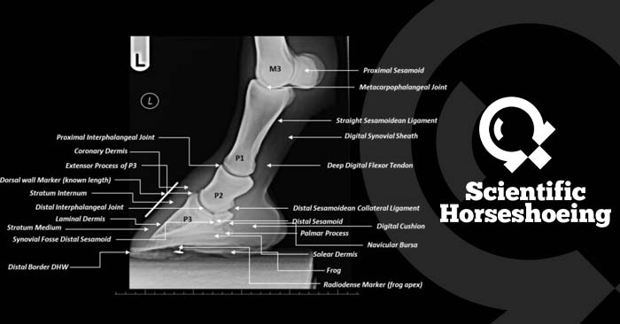
Diagnostic Imaging For Farriers
£9.99 Plus VAT
Lameness is one of the most important clinical abnormalities in horses – both in frequency and in economic impact.
Radiography is often the first method of diagnostic imaging used in the evaluation of lameness. Because of the technological evolution of digital radiographic imaging during the last decade the in the field use of this form of diagnostic imaging has widely increased. For this reason, it is now very common for farriers to request x-rays to measure themselves in everyday practice.
Whilst the farrier is not allowed to expose a diagnostic sentence but is supposed to be capable in reading and understanding x-rays to understand shoeing prescriptions and their reasons as well as to understand their achieved or not achieved result. Detailed radiographic evaluation of the horse’s foot is very useful in preplanning and management of trimming and shoeing of a horse’s foot and in the prevention of lameness. Horse’s exhibiting poor foot conformation, imbalance, or abnormal patterns of growth can be clues to impending foot disease and lameness. Radiographic evaluation, alongside a thorough clinical evaluation supported by in depth anatomical knowledge, of a horse’s foot gives tremendous insight into the relationship between the structures within the foot and between the foot and distal limb.
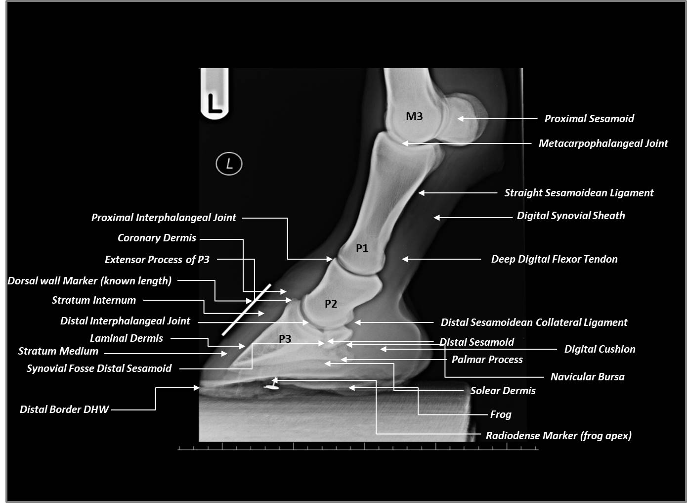
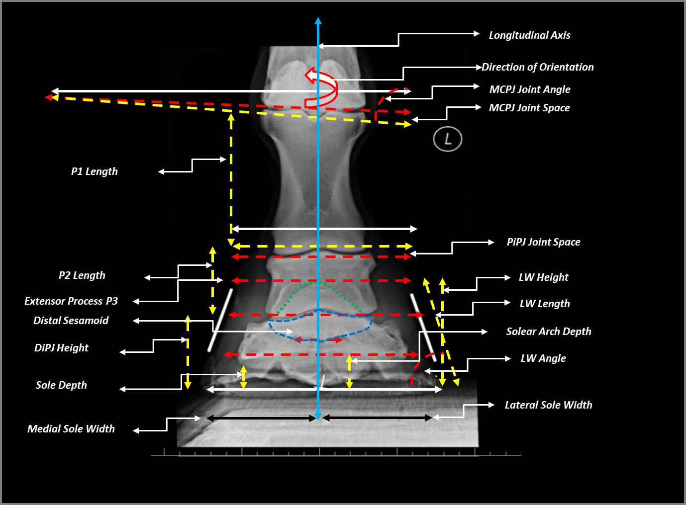
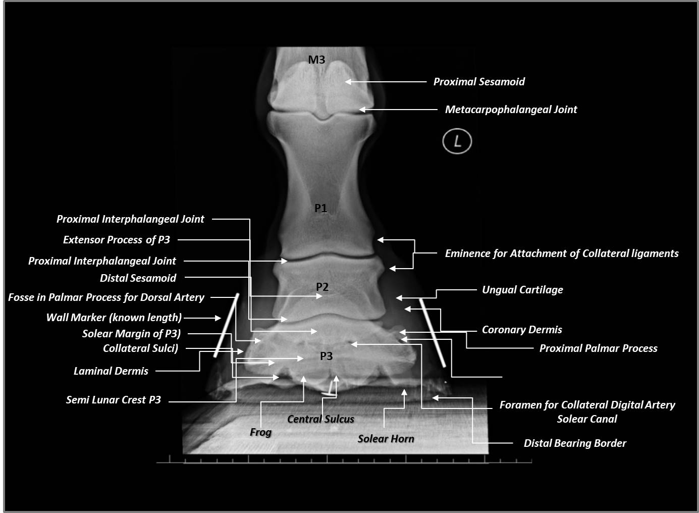
To achieve the goals of ameliorating lameness by the restoration of biomechanical efficiency farriers need to adopt a systematic approach when assessing radiographs. This approach must be based on a comparative analysis between that which is currently considered normal and the images from a planned radiographic study of the foot. For this reason, it is essential that farriers are familiar with, not only the range of radiographic views, but radiographic identification of anatomical structures and what is considered normal for any projected image.
This brief chapter aims to be an instrument for the farrier in reading x-rays, to become capable to read them and use them relatively to farriery purposes. The most radiographed areas encountered by farriers are Feet, Fetlocks (metacarpo/tarso phalangeal joints), Carpi, and Tarsi & Stifles


