Scientific Horseshoeing logo
The art and science of horse shoeing

The Art and Science of Horse Shoeing

by | Apr 29, 2022 | 0 comments

Introduction:
The purpose of this text is to focus on the most useful knowledge for formulating the principles and objectives of hoof care and shoeing. It is intended to enhance the awareness and practice of all equine professionals, as well as owners and riders. It is hoped that this text represents a thorough orientation in how to trim and shoe horses taking a “why and what for” approach to the subject whilst helping students of the subject to accomplish the objectives of the theory presented here by enhancing the understanding of the mechanics of trimming of hooves, fitting of shoes,

Background
The conformation of the equine hoof is considered an important factor affecting performance of the horse (Linford, 1993). Poor hoof conformation is a consequence of the anatomy of the horse and biomechanical function in high-performance activities and has been linked to risk of injury in horses (Kane et al., 1998). The equine hoof serves as the interface between the ground and the skeleton of the equine limb; its structure is capable of dissipating large forces associated with impact shock and loading. Hoof care professionals claim that the correct foot balance is critical in maintaining health and biomechanical efficiency (Johnston & Back, 2006), but the actual dimensions of the ideal hoof model have not yet been clearly defined. During the last century various models of hoof trimming and correct hoof balance, largely based on the historical works of Russell (1897) and others (Dollar & Wheatley, 1898) have been debated, yet to date there are little in the way of scientific data and agreement on the optimal model of hoof conformation (Thomason, 2007). Hoof conformation can be altered by human intervention, such as hoof trimming and the application of horseshoes (Kummer et al., 2006; van Heel et al., 2005).

Soundness in the equine foot relies on the three primary optimal functions of the foot—impact, all affect the health of the horse, and all three need to be considered concurrently in diagnosing and treating foot disease. Both veterinary surgeons and farriers have observed that high stress and displacements of the hoof capsule, such as flares, dishes, rings, cracks, and imbalances which often result in pain and discomfort. Recent scientific investigations offer an improved understanding of the three functions of the hoof and can advance the farriery treatment of horses as well as improve the ability to formulate trimming and shoeing plans for farriers, who are often responsible for any foot related treatment of the diagnoses. However, empirical observation, personal experience, and pragmatism have sustained the activities of trimming and shoeing for thousands of years. Factors surrounding biomechanical dysfunction of the equine hoof and the relationship with balance and morphology have perhaps not been the focus for rigorous scientific investigation. By investigating these factors there is the potential to inform and influence equine hoof care, with the ultimate aim of preventing or limiting the likelihood of injury and disease in the equine hoof.

The art and science of horse shoeing
Farriery must be studied in the same way as any other science subject using evidence based knowledge and understanding to maximize the duration of the horses successful working life within the limits of that animals individual conformation. In order that a farrier may become successful in practice this often necessitates a compromise between the need to maintain an animal in work and the need to maintain the natural physiological function of the foot. Shoeing is both an art and a science, and by making use of this study the farrier may communicate with veterinarians, horse owners and other farriers, using terminology which is readily understood. Clear communication in this way will assist the farrier to more correctly shoe or treat the horse and to gain the maximum benefit when presented with articles, papers and books.

A sound understanding of the nature and function of the horse (and in particular the lower limbs) inspires confidence in the farrier and so will greater profit his business and the economic wellbeing of both the horse and its owner.

A history of shoeing horses
There have been different opinions expressed on the origin of the horseshoe. Some historians have credited the Druids, although there is no hard evidence to support this claim, as the first to use iron shoes as a preventative measure against excessive hoof wear. Written records describing the use of nailed shoes are relatively late, first appearing around AD 900. There is very little evidence to suggest the existence of nailed-on shoes prior to AD 500 or 600, although there are archaeological examples: a horseshoe, complete with nails, dating to the 5th century A.D. has been discovered and evidence suggests that around 1000 AD, cast bronze horseshoes with nail holes became common in Europe. Commonly the design consisted of a scalloped outer rim and six nail holes.

By the time of the Crusades (1096–1270), horseshoes were widespread and frequently mentioned in various written sources (Encyclopædia Britannica, 2005). By the 13th century, shoes were forged in large quantities and could be bought ready-made. Hot shoeing, the process of shaping a heated horseshoe immediately before placing it on the horse, became common in the 16th century and in 1751 Bridges wrote his treatise titled “No Foot, No Horse” on the proper care and maintenance of hooves, a term in continued use to this day.

historical 10th century british horseshoe

Basic Principles of good horseshoeing

The aims and objectives of this post are to stimulate debate on the fundamentals of farriery in the search of trimming and shoeing protocols that account for the individual biomechanical variation within the population, highlight the consequences that individual conformation might have on form and function of the foot. In doing so, the work will examine the role that science has played in the understanding of the dynamics of the foot under load and the role of mechanical forces on the morphology of the hoof whilst discussing trimming and shoeing methodologies that might manipulate those forces for a biomechanically more efficient model.

Empirical observation, personal experience, and pragmatism have sustained the activities of trimming and shoeing for thousands of years. Factors surrounding biomechanical dysfunction of the equine hoof and the relationship with foot balance theories and there application with hoof morphology has perhaps not been the focus for rigorous scientific investigation. By investigating these factors there is the potential to inform and influence equine hoof care, with the ultimate aim of preventing or limiting the likelihood of injury and disease in the equine hoof. To better understand the relationships this work will briefly set out the background to currently accepted hoof care and horseshoeing practices. However, any review of these practices would not be complete without an elementary discussion on the anatomy and physiology of the limbs and feet. Such a discussion is indispensable as it enables a better understanding of the relationship of the hoof to the dynamics of movement and the foot and limbs interaction with the environment and substrate. Inevitably, the interaction between the foots bearing border and the ground are governed by fundamental laws of physics, it is essential to be aware that the practical interpretation of these laws dictate the basis of analysis of evidence used to formulate the major concepts of this work

Shoeing horses has often been termed a “necessary evil.” Certainly it is not natural, but neither is the domesticated life most horses must endure. Domestication results in a separation of the horse from the natural environment and life style which directed its evolution. The “unnatural” conditions of domestication usually include wet climates, restricted activity, changes in nutrition and breeding factors, often imbalanced human “training” and, of course, trimming and shoeing. The hoof’s shape and function are direct expressions of a horse’s environment, manner of movement or locomotion, and amount and type of physical activity.

Domestication, or removal of any horses from the dry, open ranges of their evolutionary habitat and their natural, vital, socially motivated physical development, promotes potentially damaging changes in healthy hoof shape and function. Such changes in hoof shape and function can obstruct movement and imbalance weight distribution in the legs, thus leading to restricted performance or lameness in the horse. The distinctions between the natural and unnatural characteristics of hoof shape are important. And so too becomes the necessity to practice hoof trimming and shoeing in ways which promote natural and discourage unnatural hoof shape. Similarly distinctions between balanced and imbalanced locomotive behavior also become important in understanding the origins and effects of unnatural hoof shape.

The acts of trimming and shoeing only become “evil” when they fail to compensate for unnatural influences or directly promote unnatural and thus unhealthy hoof shape and function. In order to best serve the interests of the horse, and thus the horse owner, hoof care and shoeing must be founded in:

Intimate knowledge of what the structures of hoof and limb are: their anatomy and histology
 Understanding of how those structures function together: their physiology and the balanced locomotion of the body it can produce under favorable conditions.
 Understanding of how the above aspects and functions relate to the horse’s “evolutionary design;” both the overall development of the body and the specific shape of the hooves which contribute to the most natural or efficient, sound locomotion.
 Analytical attitude that can discriminate the intricate factors involved in the complex relations of conformational and locomotive characteristics of individual horses which are dramatically exaggerated by the variable conditions of domestic environment and activities.

The historical reasons for shoeing horses
At this juncture it is worth remembering why we shoe horses. There are 5 main objectives and these are summarised in the basic functions of shoeing and hoof care. These five objectives are accomplished primarily by hoof shaping and shoeing combined with adjustments in the horse’s environment and training which promote relatively balanced or efficient locomotion.

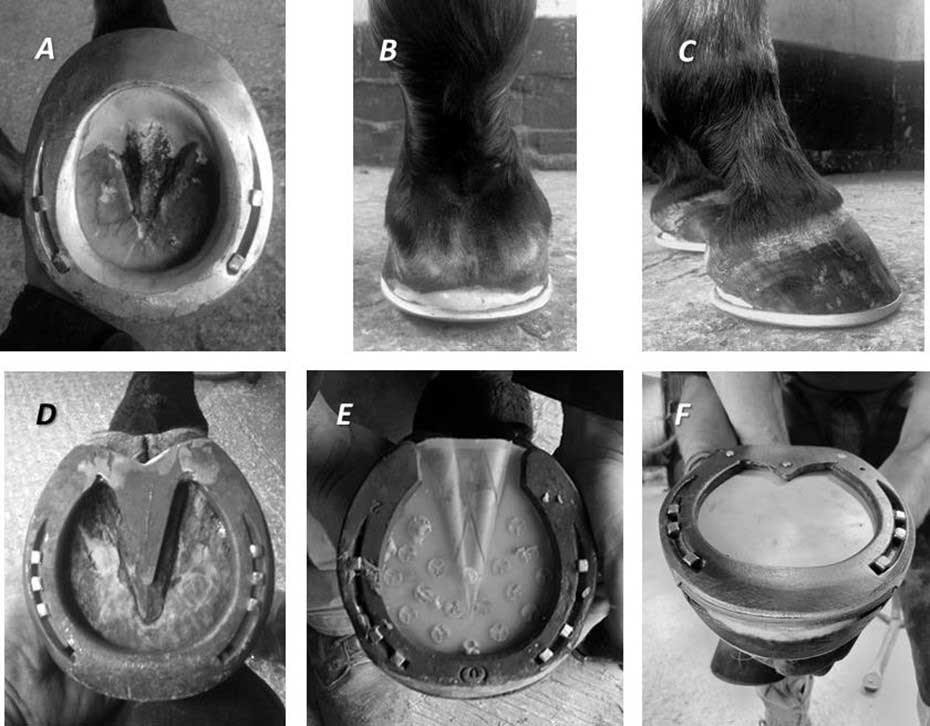
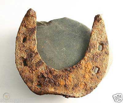
1. Maintaining the shape of the hoof- Hoof care professionals insist that correct foot balance is critical in maintaining health and biomechanical efficiency (Johnston & Back, 2006) but the actual dimensions of the ideal hoof model have not yet been clearly defined. The debate over the correct or desired proportions and angles associated with a ‘normal’ hoof capsule and what might constitute a balanced foot has been a source of contention for farriers and hoof care professionals over many years. The focus of current farriery teaching is based on maintaining correct geometric hoof balance. It is believed that geometric balance to a prescribed model promotes the most efficient form and physiological function within the foot and limb and therefore limits injury and disease to the foot and lower limb (Butler, 2005). When discussing balance, as it relates to the equine distal limb, however, the terms conformation and foot balance are often used interchangeably; more accurately conformation describes the size and shape of the musculoskeletal structures and the way in which they are spatially arranged. Foot balance though describes the way in which the hoof capsule relates to all the musculoskeletal structures of the limb. To understand the basis of foot balance in the horse a detailed understanding of anatomical form and function is required.

Good hoof care must simulate the effects of the natural influences which directed the evolution of the hoof to produce its optimum healthy shape and function. The equine hoof encapsulates and protects the bones and sensitive structures of the distal limb. The outer hoof capsule grows distally from the proximal border to the bearing border and is generally in balance with the amount of wear that naturally occurs as the horse travels over the ground (Pollitt, 1990). The growth rate of the hoof wall has been estimated at 7mm every 28 days taking on average 9 to 12 months for a hoof wall to renew itself (Pollitt, 1990). Domestication and continued work on abrasive terrain can compromise the delicate balance between growth and wear and may lead to lameness with economic implications associated with loss of animal performance, historically; this necessitated the need for professional foot care and protection in the form of a shoe.

2. Protecting Hoof and Limb- The hoof and thus the limb and horse, must be protected from damage, injury, excessive wear, deformity and disease in order to preserve or restore healthy shape and function.

Design criteria of the shoeing plan, irrespective of fitting style, must have at its core the principle of minimizing the unintended consequences on the normal anatomical and physiological function of the lower limb and foot. Within the limits of the activity that the horse is to undertake the application of shoes should as far as practically possible maintain maximum biomechanical efficiency whilst enhancing performance and grip and preventing strain related injuries. Science has shown that physiological health of the foot is best served where all the epidermal structures are engaged in appropriate weight sharing during the stance phase (Hood et al 1997; Clayton 2011). Once attached, and by whatever means, the shoe becomes an integral part of the limbs propulsive and load bearing mechanism and as such careful consideration needs to be paid to the material selection for the shoe and how that might affect the foots interaction with the terrain or surface the horse normally works on, method of attachment and the position and nature of performance enhancing ancillary features. All will directly affect the mechanical efficiency of the whole horse. Every shoe that is applied will have unintended consequences. Careful consideration to the most effective way of mitigating the unintended consequence is a vital aspect of the shoeing plan in order to maintain the general health and function of the foot. Most importantly however differences in performance horse shoeing styles will necessitate variations in the shoeing cycle.

In order to discuss the design of a comprehensive shoeing plan, it is necessary to assesses criteria for shoeing. It is essential that a common interpretation of the anticipated use of the horse is agreed and understood by all those involved in the management, welfare and training of the horse. Terminology commonly used in farriery can often be interpreted differently, for example use the description hunter in the UK and a shoeing style based on a peripheral outline fit designed to minimize premature shoe loss and injury will be adopted by the farrier with the recommendation for a much reduced shoeing cycle. The act of farriery has been described as both art and science which has a direct influence on function of the structures within the foot. It is the author’s belief that a sound trimming technique based on anatomy and physiology of the foot is the mainstay of farriery intervention. The foot’s ability to maintain its integrity and health to from trimming alone will be dependent on the integrity of those structures and the mass or density of the foot present. Various shoes are then applied to protect or compliment what has been trimmed and to manipulate the basic biomechanical forces acting on the foot.


The art and science of horse shoeing
Figure 2 this is a good example of a typical English hunter front shoe fit style. This style of shoeing is designed for horses working at high speed over mixed terrain. The heels of the foot are reduced in height so as to extend the bearing border palmar/plantar with the heels of the shoe dressed and fitted to correspond to the shape and angle of the heel buttress so as to minimize premature shoe loss and injury. The overall fit is described as a peripheral outline fit and would normally be employed in conjunction with a reduced shoeing cycle of between 21 – 28 days

The authors approach is to employ a range of shoeing styles and techniques designed to increase the ground surface of the foot, unload areas of the foot, effect or reposition breakover, and dampen concussion to some degree. Biomechanically, these techniques can: A) change dorsopalmar or mediolateral orientation of the foot thus changing forces by moving CoP (Hagen 2016), B) reduce the forces associated with breakover (Moleman et al 2006), and C) decrease the stresses associated with the soft tissue pathologies by transferring load (Denoix et al 2001). Selection of a broad light weight material as possible, that will last the duration of the expected shoeing cycle, normally between 35 & 42 days, will minimize the energy requirements musculoskeletal system and effect a smoother transition of the individual limbs into the swing phase of the stride. Whilst the careful selection of the materials profile suitable for the intended terrain or surface will enhance the safety of both horses and rider.

3. Facilitating Locomotion- The movement of the horse should be assisted or enhanced (not obstructed) to promote athletic performance and diminish chances of injury.

The modem equine working environment necessitates consideration for the health and safety of horse and rider / driver / handler. This often necessitates compromises of foot welfare in the form of shoeing styles to maximize traction for different surfaces. The surfaces on which different working, leisure and sporting activities are practiced do not afford the same traction as dirt roads; therefore there is often a need to add to the horse’s foot some form of anti – slip device. Traction can be improved through the use of different shoe profiles or the addition of artificial ground surface projections such as studs or caulkins. However, these devices affect the delicate biomechanical relationship of the internal anatomical structures of the foot which can in turn result in increased strain of both the axial and appendicular musculoskeletal system increasing the likelihood of either interference injuries through changes in gait or repetitive strain injuries or pathologies which result in premature lameness.

4. Compensating for and managing abnormalities- Weaknesses and deformities in hoof, limb or locomotion which may cause and/or result from injury should be compensated by trimming, shoeing or training when possible. Excessive or unnatural wear can often be related to excessive or abnormal weight bearing or to loss of flexion of limb during motion for example dragging of the toe as the foot lands or as it leaves the ground (break-over). There may be a number of causes including limb and foot conformation as well as a response to pain and pathology leading to self-evident changes in the foots biomechanical interaction with the ground. For example this is often witnessed in cases of navicular lameness and bone spavin disease which frequently result in excessive wear of the dorsodistal hoof wall and a loss of solar mass. Farriery management of abnormal wear of either the hoof or shoe is often achieved by simple manipulations of the shoe such as rolling the toe of the shoe to mirror the appearance of the worn bearing surface of the hoof.

5. Provide Therapy- Causes of injury or lameness related to hoof shape and function or locomotive actions of the limb should be eliminated or alleviated when possible.

To evaluate the affects therapeutic shoeing interventions, it is necessary to define the term therapeutic farriery. Therapeutic farriery could be described as “the art and science of affecting / influencing the structures of the foot”. This is primarily achieved through the manipulation of forces to affect a mechanical advantage which will result in the relief of pain through the transfer of load, the provision of support or providing stability of off axis movement to reduce compressive or tensile strain. Often therapeutic intervention takes the form of prescriptive horseshoes for specific conditions such as the heart-bar shoe for laminitis or the egg-bar shoe for navicular disease to name but a few. In reality the myriad of these types of shoes actually offer the farrier a number of variants that can be added to standard shoes to be applied in the manner previously described. In each case these variants should be carefully selected to match the desired effect to ameliorate individual symptoms with careful consideration given to mitigating the unintended consequences and particular risks to the wellbeing and function of the foot and limb as a whole. Proper trimming is paramount to the recovery of the foot and is more important than the type of device applied to the foot. The hoof must be trimmed with the goals of establishing a normal angle, balance, and length of the foot, and eliminating diseased or distorted horn. 

Proper identification and analysis of problem areas, like hoof capsule distortion, enables farriers to implement changes to landing strategies with the aim of reducing the effect of landing force by increasing the surface area and thus minimize injury. In biomechanical terms forces responsible for the stress directed over the foot cannot be eliminated. However their direction and duration can be manipulated to relieve strain. In simple terms farriery interventions can manipulate force (mass x acceleration) by creating a pushing or pulling action on the point of force trajectory. This can be achieved by adaptation of the shoe. The addition of material to the bearing surface area effectively pulls the force towards the intervention whilst the removal of ground bearing area pushes the force away from that area.

An important principle of any therapeutic shoeing plan is to dissipate load over as large an area as possible by shoeing the foot full through the heels and extending the bearing border palmarly. Historically, egg bar shoes and variants have been used for this purpose; however, the degree of extension can create a lever effect concentrating load on the edge of the heel (O’Grady 2011). Straight bar shoes, heart bar shoes, onion shoes, roller motion shoes or other wide-webbed heel devices have been used successfully to support the heels by manipulating forces across the foot. Implementation of these is however often considered impractical for long-term use in a competing athlete. The addition of solar support in the form of pour-in pads, impression material and or various types of proprietary pads can contribute to increased stability and load sharing whilst often providing temporary relief of clinical symptoms and aiding the prevention of any further reduction in integral strength of the hoof structures.

Therapeutic shoeing plans aim to (1) reduce acute pain and inflammation, (2) optimize repair of the injured tendon or ligament, (3) to reduce the effect of adverse biomechanical forces acting on the injured structure. This is primarily achieved by improving the dorsopalmar/plantar and mediolateral orientation of the foot and lower limb through trimming and the attachment of shoes designed to manipulate force and compensate for conformational anomalies. These types of modified horseshoes are primarily thought to facilitate lateral breakover during turns by reducing torque subsequently relieving compressive and tensile forces in and around the joints and the connective structures (Denoix et al 2007). A variety of horseshoes have developed with the common purpose of optimizing biomechanical efficiency of the foot and to relieve repetitive strain such as that experienced by the collateral ligaments and for the treatment of articular disorders (Gregory 2007). These types of modified horseshoes can alter the height (Side Wedge shoe), the weight bearing surface (Wide Branch or Wide Toe shoe) and/or the torsional forces experienced at breakover (Roller shoes). All can influence the biomechanics of the distal limb during stance and locomotion (Denoix et al 2007). However the effect of these types of horseshoe modifications are often surface dependent and indeed their application may be contraindicated in certain environmental and management circumstances. The unintended side effects and the individual duration the horses can tolerate them may indicate a temporary limit for the application of any modified therapeutic horseshoe. These types of therapeutic shoes are not designed to be used all of the time. The author believes however they may also have a useful role to play in the prevention of soft tissue injury where severe foot imbalance and conformational abnormalities are present.

However, the effect of these types of horseshoe modifications are often surface dependent and indeed their application may be contraindicated in certain environmental and management circumstances. The unintended side effects and the individual duration the horses can tolerate them may indicate a temporary limit for the application of any modified therapeutic horseshoe. These types of therapeutic shoes are not designed to be used all of the time. The author believes however they may also have a useful role to play in the prevention of soft tissue injury where severe foot imbalance and conformational abnormalities are present. When applied for the treatment of a specific injury, it is crucial to keep in mind that relieving certain structures of the distal equine limb immediately causes more stress on the antagonistic structures and may lead to subsequent pathologies. These shoes increase biomechanical stresses on other supporting structures of the limb, which can lead to failure of those structures (Richter 2015). The shoes are meant for use while an injury is healing and during a portion of the rehabilitation period, in general for less than 1 year. The ultimate goal is to return the horse to use of a plain shoe at the earliest opportunity. This requires that these types of shoe are adjusted as the injury responds to treatment. Often during this time the foot will change, it is essential the farrier ensures that the foot remains well balanced post treatment maintaining a foot conformation that prevents an increase in stress on the formerly injured structure.

the art and science of horseshoeing
Figure 3 examples of common therapeutic variants commonly used for dissipating load by increasing the surface area and providing support and stability to internal foot structures through the sole and frog whilst relieving compressive and tensile forces on the DHW. A) An egg bar shoe with leverage reduction, the palmar aspects of the foot are engaged in support with the addition of impression material. B & C) The correct fitting of palmar aspect of the egg-bar shoe. The outer radius should mirror the coronary band and terminate adjacent to the heel bulb. D) A steel frog shaped insert engages the frog into a weight sharing role whilst reducing the direct load on the heel buttresses. E) A full sole pad with an inverted frog shaped piece which is normally fitted so that the frog piece is positioned parallel to the bearing border of the shoe engages the frog in weight sharing during the stance phase of the stride. The addition of 20 shore silicone pour in pad material engages the sole in weight sharing without excessive sole pressure. F) A traditional straight bar shoe provides stability to the heel area relieving the outer border of the ground surface reduces shear force during unrollement in this case a leather pad and silicone pad are used to engage the sole in weight sharing (Photographs A – C courtesy of L. Bentham DWCF Photograph F courtesy of C. Montagna).
standard UK concave riding horse style
Figure 4 A good example of standard UK concave riding horse style horseshoeing
This paper describes a method of treating cases of unilateral palmar/plantar laminitis using the Steward Clog ®(aka Wooden Shoe). This method varies significantly from a previously advocated technique using the Wooden Shoe. In this paper we report on the use of a different/modified technique to load the unaffected wall in horses with marked unilateral displacement (including sheared heel) of the distal phalanx. Additionally, the EVA/Wood Clog used- represents further development of the shoe as originally described (Steward Clog 2.0®).
Introduction:

The variety of benefits the Steward Clog® (Wooden Shoe) offers the laminitic horse is evident by the widespread usage throughout the horse industry. Ease of application and design modifications allow the shoe to be utilized effectively by practitioner and/or farrier without an expertise in therapeutic shoeing. The application process allows a non-traumatic procedure that relies heavily on the input of the horse in maximizing the horse’s comfort. Lateral and dorsopalmar (DP) radiographs are vital information to aid the proper trim in the therapeutic shoeing procedure.

Unilateral distal displacement can be accompanied by dorsal capsular displacement, in complicated laminitic cases. The damaged lamellae allow overload injury to portions of the wall / bone interface and this can manifest itself in variable displacement of the distal phalanx within the hoof capsule. The mechanical pull of the deep digital flexor tendon, combined with the weight forces produce the most common manifestation of laminitis- dorsal capsular displacement/phalangeal displacement. The pattern of displacement varies with the distribution of damage and load, the exact pathophysiology of which has yet to be determined. Displacement of a particular portion of the third phalanx is usually due to the area of displacement being overloaded and the damaged Suspensory Lamellar Apparatus’ (SLA) inability to support the load. The classic manifestations of laminitis are usually manifested as overload injury of a particular area of the distal phalanx. Depending on the extent of lamellar damage and the particular amount of load to P3, the variable displacement of the third phalanx is indicative of amount of damage and amount of load causing shear forces to the SLA.

Unilateral displacement is usually medial in the front limbs and lateral (author’s opinion) in the hind limbs (see Fig.1) -in typical cases. Conformational imbalanced (valgus) overloading of the medial wall’s damaged lamellae (in the fore limbs- with non-ambulatory perfusion deficits) is thought to be the usual cause of fore limb’s medial displacement (see Fig. 2). The hind limb is (typically) imbalanced overloaded on the lateral aspect of the hoof (because of the single leg resting stance) and this can often be manifested as lateral displacement (if the lamellae have been sufficiently damaged and overloaded). The diagnosis is based on the physical appearance of the foot, asymmetrical pain distribution and radiographs. Dorsopalmar (DP) radiographs are very helpful in revealing the condition. Abscesses and other pathologies causing particular areas of vascular discrepancies can alter the common areas of manifestations of this pathology. Current dynamic Positron Emission Tomography (Florodeoxyglucose) scans (Andrew VanEps, DVM) have shown extensive areas of (ie,-medial) wall that have deficits in vascular perfusion when non-ambulatory- in clinically normal feet. These deficits resolve upon ambulation. This may account for the medial UPD when dorsal phalangeal laminitis occurs, for example. The non-ambulatory lesions become pathological when the patient suffers lamellae damage and the painful condition further diminishes the compromised SLA wall perfusion.

In cases of dorsal phalangeal rotation of the third phalanx, the anterior ½ of the coffin bone distal displaces around the second phalanx’s distal condyles. The caudal ½ of the coffin bone displaces upward (unless a sinker). The lamellae in the heel area are stretched and are receptive to weight load, which would apply forces to aid in realigning the lamellae as the weight forces the bony column (P3) distally and the ground reaction forces (GRF) produce an upward vector force on the frog area of the hoof. When the lamellae are sufficiently damaged and overloaded, shearing of the lamellae occurs and the weight forces (WF) cause the distal migration of the affected portion of P3 (ie. – medial wing). Heel hoof growth can be considered to be enhanced because of the wide growth rings, but most of the enlarged ring is due to lamellar stretching and the pull on the newly formed hoof wall tubules in an unencumbered growing environment just below the dorsally displaced coronet. The horn tubules may be formed at the same rate around the coronet, but the stretching/compression and traffic jam/ fast-lane areas of tubular maturation and development just below the coronet account for wall formation/growth and growth ring conformation / thickness. The dorsoaxially displacing wall helps to shear the SLA as the third phalanx displaces distally. This is a tectonic plate-like biomechanical problem- comparable to an earthquake.

Once the displacement has occurred, a dip (recess) can be palpated in the integument immediately proximal to the wall on the affected side. The ungual cartilage/ wall relationship is displaced. Additionally, the toe and unaffected heel of the hoof capsule displaces dorsoaxially and rotates towards (yaw effect) the separated side (the plastic deformation usually occurs over time). This displacement (distal pitch of P3) and rotation (yaw of the hoof capsule) increase with time as the wall on the affected side shows little or no growth (due to disrupted blood supply in the damaged SLA) and the opposite side shows normal or increased growth. As the third phalanx rotates distally (pitch), the affected hoof wall is plastically displaced dorsoaxially (medial/roll (inversion roll), pitch-distally). This displacement is similar in pathophysiology to cases of “sheared heels” (traumatic/subclinical unilateral palmar laminitis (stretching) with dorsoaxial wall displacement), but the amount of overload shear trauma (and SLA damage) to produce heel (P3 &/or heel wall) displacement in the two cases- is substantially different. Both cases occur as a result of overload (shear or stretching) to the (damaged) lamellar apparatus of the particular area of the hoof. Laminitis cases have an enzymatic damage/weakening of the lamellae that lends itself to failure- if the lamellar damage is sufficient and overload exceeds this overload failure point. Unloading the affected wall is essential to the success of therapeutic intervention in both conditions. In severe cases of unilateral palmar/plantar displacement (UPD), extreme measures usually have to be implemented to have a chance of success. This involves unloading the affected wall by floating (cutting short/unloading) the affected heel, load redistribution, moment arm force reduction- to the affected lamellae, unloading pained areas, and stabilizing P3. The upward displacement of the hoof wall and distal displacing third phalanx create complicated biomechanics for resolution, as the wing of P3 is forced upward (biomechanically) as the coronet and affected wall needs to be displaced distally via plastically deforming biomechanics. The new growth attachments must be plastically deformed or artificially moved back into a more normal relationship- for functional healing to occur.

Previous descriptions¹ of the use of the Clog (wooden shoe) in the treatment of unilateral distal displacement have focused on extending the shoe towards the unaffected side to shift the center of pressure away from the most damaged lamellae, but the author finds this somewhat useful in some chronic, stable pathology, but not relevant in cases of acute laminitis. (See drawing-A.Parks)

The full roller motion design feature, particularly the mediolateral breakover (roll) portion of the Wooden Clog, and the subsequent use of an ethylene vinyl acetate (EVA) pad (similarly designed)- allowed the patients to (immediately) formulate a consistent, comfortable therapeutic formulation that enhanced soundness. This palliative effect, combined with other procedures (ie.- coronary grooving/wall resection) produced the most consistent success rate (curative effect).

Materials and Methods:

Plywood (1.125 inch) is cut and shaped to form the basic Wooden Shoe design. Mediolateral sloping can be increased to allow the patient to easily manipulate foot loading. The longer the slope (D/P) is extended toward the centerline of the shoe, the easier mediolateral (roll) breakover is achieved (see Fig. 3) and the hoof can shift weight to the less painful heel by slightly rolling the shoe- inversion roll for medial UPD and eversion roll for lateral UPD.. The sloping can be extended past the centerline to form a wedge effect to the shoe with the widest portion of the wedge under the affected heel (see Fig. 4).
The addition of EVA (ethylene vinyl acetate) to a layer of plywood (with the perimeter of the EVA cut in the same shape as the basic Wooden Shoe- Steward Clog 2.0®) allows the patient to self-adjust (plastically deform) the EVA and adds the same biomechanics to the shoe (see Fig. 5). Additional concussion absorption is a very beneficial feature- as well as the selective stabilization the elastic, plastic properties the EVA possesses.

Results:

Five horses (Four QH, 1 Arabian, ages 3 – 20 years) with bilateral medial displacement (front feet), 9 cases (8 QH, 1 Paint, ages (2-22 years) with unilateral displacement (seven front and 2 hind feet) have been treated using this technique. Increased comfort and a better radiographic DIP joint alignment were noted when modifications were made.
Successful outcome depended on the amount of lamellar damage and the amount of vascular damage as evidenced by bone loss and permanent bony displacement. Four horses (4) with minimal bilateral displacement- returned to pasture sound, six (6) patients with minimal unilateral displacement in a single foot are (occasionally) ridden at a walk. Four (4) of the horses are maintained in restricted environments with limited soundness.
Six feet required partial wall resection after displaying wall detachment (greater than 1 inch of detachment). The (dead / nutritionally compromised) wall below the attachment appears to shrink (dehydrate) toward the distal phalanx and the growth from the coronet prolapses over the distal wall if it is not removed. The wall detachment is often mis-diagnosed as a “gravel abscess” as it presents itself at the coronary band.

Discussion:

This shoe allows the patient to selectively load and self adjust breakover (pitching) and wedging (roll). The Steward Clog (aka Wooden Shoe) requires the practitioner have a knowledge of the breakover needs of the particular case: whereby, mediolateral point of breakover (roll) is appropriately applied to a nondeformable shoe material (wood, steel, aluminum, rubber, etc.). The addition of EVA to the shoe’s solar surface allows the patient to apply the point of breakover- and subsequent wedging effects to the shoe. Both shoe designs enable the horse to re-adjust the mediolateral breakover as the hoof growth’s mediolateral plastic asymmetries occur between shoeings.

The sloping of the mediolateral surface to the (EVA/Wood, Wood) Clog allows the patient to “roll” the shoe to realign the DIP joint and to load the hoof according to the comfort of the patient. The use of the EVA material on the bottom of a layer of plywood allows the patient to easily, immediately conform the shoe to the biomechanical / comfort needs of the patient (Steward Clog 2.0®). The plastic properties of the particular EVA allow the basilar surface (usually toe in typical laminitis, but medial or lateral heel in cases of UPD-both deformations may be needed in a particular case) to maintain the plastically compressed areas. All the while, the EVA maintains elastic properties to absorb concussion and further conform as the hoof grows and horse heals- or pathology worsens. These cases- immediately- loaded the shoe to the unaffected side, wedging (mediolaterally) the shoe such that the DIP joint was realigned (see Fig. 1,3,5 ) when the EVA foam was present. This is analogous to how a person would walk if the suffered a cut to the medial heel of their foot. One would roll up (dorsodistally pitch) off the heel (inversion), load the lateral portion of the foot (heel) and place the foot towards the midline (axially) to walk (redistribute load). The base slopes of the shoe can be altered to redistribute load. The affected side’s basal shoe heel/wall/ toe solar edge can be beveled at a 35 degree and the unaffected (lateral shoe branch) side can have a lesser slope to the ground connected surface (0 to 15 degrees). This will transfer more load (GRF/WF) to the lateral wall.
The basal (ground) surface of the Wood Clog (or urethane Clog) can be modified by solar grinding or addition of a wedged pad to produce an inversion roll (wedge sloped laterally). This will painfully overload the medial wall; therefore, wall floating measures should be implemented and load redistributed to offload the affected wall’s/solar load share. The dorsal surface of the “wooden shoe” should be ground down starting at the second nail hole and gradually slope the wall and solar area caudally to the affected heel. The triangular shaped area is from the wall to the medial sulcus of the frog. The surface of the shoe will be removed at a continual deepening slope to unload the floating wall. The wall can be trimmed in a sloping fashion (opposite the sole recess) to insure wall contact is avoided. The dorsoaxially displaced wall/coronet will usually migrate distally- to return to a more normal position – if adequately unloaded. Another option is to construct a W shaped caudal pad to redistribute the load off the medial wall (see Fig.7). Leather or wood can be used to construct the W pad. It is extended dorsally to the quarters. The pad(s) are under the shoe, and extend to the caudal region to provide wall and frog support. The affected side can be reduced by grinding the shoe surface of the pad in the above described fashion from the 2nd nail hole to the heel. The recessed area will extend under the bars and extend to the medial sulci of the frog. The recess should extend forward to unload the area of the sole that would be occupied by the distally projected shadow of the wing of the P3. The central leg of the W is to support the frog. The pad can be expanded in the frog area to accept more load by adding a layer of SIM to the central and lateral sulci of the frog. A configured pad (thickened frog) can be used to make sure the frog is actively loaded to overload the frog and make sure the medial wall and sole are protected (unloaded). Wetting a leather pad insures proper fit once the pad is applied and allows easier plastic deformation- via hammering. The pad can be conformed (hammered) on an anvil – (much the same as steel) to thin the medial wall/solar area- from the second hole, to the medial sulci of the frog and caudally to the heels ( wall to frog sulci / quarter to heel recess). This orthotic package (purchase) can be shod with a normal shoe that has the medial branch unloaded as described above. This is useful for a sheared heel that must continue working. A (Z bar) shoe can be used to transfer load to the lateral wall in some cases. The manipulative mixing, curing of the sole impression material can offload the affected heel and overload the unaffected heel. For example, the Shore value of SIM can be softened by 50% by using ½ the white portion of the 2 part SIM in the mix. This will reduce the Shore value by 50% and soften the SIM, effectively unloading pained areas. Curing/ manipulative adjustments can be used to overload normal areas of the frog/unaffected heel. Loading the hoof just prior to the SIM setting up, and not fully loading the leg will allow the unaffected area to be (actively) overloaded- increasing the area’s share of WF.

Excessive damage to the affected wall can result in an (comparatively) increase in hoof growth on the unaffected side as compared to the affected side. This causes increased wedging (pitch-d/p- and roll m/l) to the hoof capsule between trims (see Fig. 5). The affected wall (or portion) has lost a large portion (or all) of the nutritional and physical support of the SLA. This can cause a rotational hoof capsular deformity (yaw effect), over time, as the affected heel dehydrates and shrinks. Deleterious wall compression and detachment needs to be aggressively dealt with. Coronary grooving and other wall sculpting, floating methods (resections) must be done to allow the displaced wall and prolapsed coronet to return to a more normal form to re-establish a more normal function. These wall growth distortions cause a force on the elastic/plastic hoof capsule that results in the plastic yaw motion/deformity to be toward the affected side,. The faster growing un(less)affected side adds substantial mass to the lateral wall (in cases of medial sinkers) and further adds to the medial yaw effect. The dorsal capsular rotation of P3, especially if severe (incrementally -degreed), confounds the yaw effect. The rapid growing (net vector effect) heel causes a medial yaw and the slow growing toe region produces an initial, distally- orientated plane of growth (just distal to coronet) as it seeks the distal extensor process (SLA) of P3. The dorsoaxially displaced proximal wall and coronet compromises (via stretching or compression) the coronary vascular and needs to be encouraged to plastically return to a more normal architecture to return a more normally functioning wall. This encourages the mitotic area to restore the new portion (section) of connecting tubules as they encounter the previously produced, immature, (coronary band- terminal papillae produced/ very pliable) wall tubules, thus, distortions of growth rings occur. These distortions are a function of the amount of rotation that P3 has suffered. Their growth is retarded by coronary band vascular compression/stretching- because of P3’s displacement,’ thus denying them of the normal nutrition of the unaffected heel lamellar circulation. As the unkeratinized (uncemented, pliable, connected) terminal tubules (continuously) exit from their coronary papillar finger-like factories, they normally are attached (easily) to the SLA (suspensory lamellae apparatus).

The normal wall tubule is displaced distally as it is seeks to be attached to the SLA- in a complex manner- as to allow a ratcheting growth pattern of the continuous tubule/lamellae complex. The tubules are responsible for the observed growth rings of the hoof. In their pliable state, as they exit the coronet (papillae factory), they are easily deformed. The wall tubules are similar to hair shafts and the collective intercemented- collection of tubules- is the hoof capsule. They are interconnected to each other to form the continuous, complex wall -with a (normally) very tough hoof wall cementing system, -after their detoured route caused by P3 displacement. They can encounter previous (slow) growth which causes a “traffic jam” and results in very compacted, small growth rings, or the tubule can be folded resembling an accordion or “pleats”(displacement dependent). In severe P3 rotation (greater than 25 degrees), the large pleated sheets of (horizontally connected) tubules can appear as “folded layers” as they form unstable, distorted hoof wall. The interconnected (horizontally) tubules are connected to the (individual) continuous (coronet to ground) hoof capsular tubules as the individual tubules follow their previously produced segments to the ground. The keratinazation and “cementation” occur (only) within the perimeters of their close proximity to the lamellar corium (SLA). It is damaged by the displacement of P3, and often does not return to an acceptable / functional state, thus distorting normal wall formation and growth.

Unilateral palmar displacement is a very difficult, devastating overload injury / manifestation to cases of complicated dorsal capsular/ phylangeal displacement laminitis. The amount of damage to the entire lamellar interface¹ combined with the amount of load to a particular area of the lamellar interface accounts for the various manifestations of laminitis. Reducing load to the lamellae- prior to devastating overload injury- is possible in some cases, but unfortunately, some cases experience devastating lamellar damage and overload injury / consequences- such that therapeutic biomechanical manipulation aimed at lessening / redistributing the load is of no benefit in re-establishing acceptable long-term soundness.

The aggressive use of coronary grooving / affected wall resection and unloading the affected wall- by trimming to allow the coronet to unload and return to a normal position -is highly recommended by the author. Any laminitic episode in the palmar (plantar) area complicates healing due to the blood supply (hemodynamics) and load bearing features generally required by the heels.

Fig. 1. a,- Illustrated radiograph showing the radiographic changes on the DP in UPD, This hind limb suffered lateral (left) displacement of the distal phalanx as evident in this dorsopalmar radiograph (b.). The increased distance in the DIP lateral joint space (white line), increased thickness of the lateral hoof wall, and differential lines of the nutrient foramen of P3 compared to the distal aspects of P2- all suggest unilateral diplacement (a., b.).

Dr. Andy Parks’ sketch of the laterally placed shoe on this depiction of a medial sinker that was described the AAEP 2007 paper- How to treat 3 Manifestations of Lamimitis using the Wooden Shoe (O’Grady, Steward, & Parks).

Fig. 2. This acute laminitis case was suffering pre-existing mediolateral imbalance that was possibly creating increased loading to the medial lamellar interface. Both front feet of this bilateral valgus deviated horse were shod similarly to the above radiograph. Wooden shoes were applied and this hoof required medial hoof wall resection 45 days later (see Fig. 5) due to severe wall disruption. Note the abnormal radiolucent area (lamellar interface area) on the medial aspect (arrow).

Fig. 3. The hoof in Fig. 1 was shod using the wooden shoe. No trimming was done to the hoof between shoeing. The shoe has been outlined in white lines. The arrow shows the medial slope (red line) that was extended to past the shoe’s midline after extending the shoe medially. (The slope of the red line is exaggerated for illustration purposes.) Note how the joint space has realigned in a more normal manner. This is accomplished by allowing the horse to increase load to the medial heel in this lateral displacement. Hoof placement will be abaxially in lateral displacers, axially in medial displacers.

Fig. 4. Following separation of the medial wall at the coronet, the separated hoof capsule was resected. The growth rings proximally illustrate the disparity in hoof growth between the medial and lateral wall before resection; the lateral wall markedly outgrew the compressed medial wall. Prior to application of the shoe, the coronary band was tilted down towards the medial side. The foot is twisted (medial jaw inversion roll) to the medial side (red line) due to disparity in heel wall growth and mediolateral shoe positioning. Following application of the shoe, the EVA compressed such that the medial coronet is now proximal to the lateral coronet (inversion roll).

Fig. 5. This EVA / Wooden shoe has self-adjusted to meet the biomechanical and comfort needs of this medial sinker. The lateral heel has grown more than the medial heel and the shoe can readjust for this difference. Soundness improved in the 30 days this particular shoe was worn due to hoof stabilization and wall resection. The resected wall allows the upward displaced coronet to plastically return to a more normal architectural arrangement with P3.

Fig. 6. This drawing illustrates the loading and compression of the EVA material in this unilateral displacement drawing. The mediolateral wedging effect realigns the joint space and aids in comfort of the patient. The radiograph displays the joint and shoe angles. The sole was trimmed with no mediolateral imbalance 30 days prior. Note the upward displacement of the affected coronary band.

Fig.7. This leather “W pad” is used to redistribute weight to the palmar/plantar hoof. The affected side of the UPD case can be unloaded by thinning the leather pad on the affected side by “hammering the wet leather pad on the anvil” prior to application. Other methods- such as recessing the shoe or cutting (floating) the affected wall- can be used to offloaded the pained region of the hoof. The unilateral wing of the displaced third phalanx is usually very painful and warrants offloading with less sole impression material, or a softer durometer SIM in the pained areas. Usually a combinations of offloading are done to allow the new wall to regain a normal SLA attachment. Photo B. The affected side can be ground down to offload the sore solear area- the entire area from the quarter to the heel if needed.

Fig. 8 (a. b. c.) These pictures are of a healed medial UPD case that must be trimmed to offset the displaced medial wall (with chronic growth disparity). The upward shift of the medial wall and the distal displacement of the medial wing of P3 create trimming complexities and are distorted between trims due to wall growth disparities.

Fig. 9. (a. b. c.) These pictures show the proper trim and shoeing prescription provided by using an EVA Clog placed on the hoof. The conforming Clog plastically deforms to the special needs of these cases. Note the level coronet in picture a. (verses the same foot in Fig. 9a.) provided by the lateral compression of the Clog. The growth of the lateral wall promotes a medial jaw as the hoof grows. Shoeing allows the hoof to alter the hoof’s inversion roll as the lateral wall grows faster than the medial wall. The dorsal capsular displacement (pitch) is complicated by the medial unilateral palmar displacement (inversion roll) and will require special trims and shoeings for the life of the horse.

Acknowledgments:

i. Declaration of ethics- No ethics of the AVMA, AAEP, or AFA were violated in applications of these methods or writing this paper

ii. Conflicts of interests- The authors received compensation for the application and sale of the orthotics in treatment of these pathologies. The Steward Clog has been used extensively since 1986.

iii, Funding/Materials/Technical Support- No funding was provided by entities/businesses or material/technical support

References:

O’Grady, S.E., Steward, M., Parks, A.H. (2007a) How to Construct and Apply the Wooden Shoe for treating Three Manifestations of Chronic Laminitis. in Proceedings. Amer. Assoc of Equine Practnrs. 53, 423-429.

Steward, ML. How to construct and apply atraumatic therapeutic shoes to treat acute or chronic laminitis in the horse. American Association of Equine Practitioners 49th Annual Convention 2003;337-346.

Parks AH. Chronic Laminitis. In: Current Therapy in Equine Medicine. 2003 pp. 520-528. Saunders, Philadelphia.

Parks AH. O’Grady, SE, Chronic laminitis: Current treatment strategies, Vet. Clinics of N. America; 19: 393-416.

Steward, M.L. The Use of the Wooden Shoe (Steward Clog) in Treating Laminitis, Veterinary Veterinary Clinics of North America: Equine Practice- Laminitis, Vol. 26, Issue 1, April 2010, pages 207-214.

Hood DM. The mechanisms and consequences of structural failure of the foot
Vet. Clin. N Am 1999; 15: 437-461.

® Steward Clog (2.0)- Well Shod Farrier Supply, 305 N. Arthur St. Amarillo, Texas 79107 (806)322-3680 www. Well-shod.com